ENCAB000AMN
Antibody against Homo sapiens ZC3H11A, Mus musculus ZC3H11A
Homo sapiens
MCF-7, GM12878, K562, HeLa-S3, HepG2
characterized to standards
Homo sapiens
A549
characterized to standards with exemption
Homo sapiens
any cell type or tissue
partially characterized
Mus musculus
any cell type or tissue
partially characterized
- Status
- released
- Source (vendor)
- Novus
- Product ID
- NB100-74650
- Lot ID
- A1
- Characterized targets
- ZC3H11A (Homo sapiens), ZC3H11A (Mus musculus)
- Host
- rabbit
- Clonality
- polyclonal
- Purification
- affinity
- Antigen description
- The immunogen recognized by this antibody maps to a region between residue 250 and 300 of human zinc finger CCCH-type containing 11A.
- External resources
Characterizations
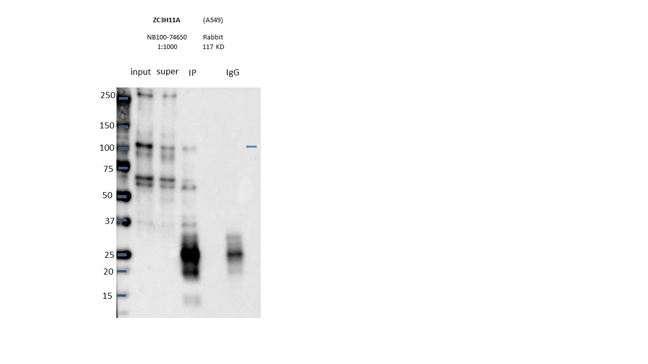
ZC3H11A (Homo sapiens)
A549
Method: immunoprecipitation
not compliant
- Caption
- Immunoprecipitation was performed on nuclear extracts from the cell line: A549, using the antibody NB100-74650. The blot shows western blot analysis of input, flowthrough, immunoprecipitate and mock immunoprecipitate using IgG.Molecular Weight: 117.0
- Reviewer comment
- Band of interest is not 50% of the overall signal in the lane
- Submitted by
- Nathaniel Watson
- Lab
- Michael Snyder, Stanford
- Grant
- U54HG006996
ZC3H11A (Homo sapiens)
Method: immunoprecipitation
not reviewed
- Caption
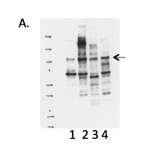
- A. Western blots on nuclear lysates from cell lines GM12878 (Lane1), K562 (Lane2), HeLaS3 (Lane3), and HepG2 (Lane4). B. Immunoprecipitation was performed on nuclear lysates from K562 cells using antibody NB100-74650 against ZC3H11A. Lane1: Nuclear lysate. Lane 2: Unbound material from immunoprecipitation with NB100-74650. Lane 3: Bound material from immunoprecipitation with NB100-74650. Lane 4: Bound material from control immunoprecipitation with rabbit IgG. Arrow indicates band of expected size (89kD) that is highly enriched in the specifically immunoprecipitated fraction. Bands indicated by * in K562 immunoprecipitate is IgG light chains. A band of ~89kD is detected by Western blotting with NB100-74650 in multiple human cell lines. Immunoprecipitation from K562 nuclear lysate efficiently enriches a single protein of ~89KD. Based on these observations, this antibody meets this ENCODE criterion.
- Submitted by
- Michael Snyder
- Lab
- Michael Snyder, Stanford
- Grant
- U54HG004558
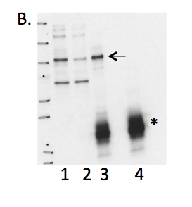
ZC3H11A (Homo sapiens)
Method: immunoprecipitation followed by mass spectrometry
not reviewed
- Caption
- Immunoprecipitation of ZC3H11A from K562 cells using NB100-74650. Lane 1: input nuclear lysate, Lane 2: material immunoprecipitated with NB100-74650, Lane 3: material immunoprecipitated using control IgG. Bands A was excised from the gel and subject to analysis by mass spectrometry. IP followed by mass spectrometry: Briefly, protein was immunoprecipitated from K562 whole cell lysates using NB100-74650, and the IP fraction was loaded on a 10% polyacrylamide gel (NuPAGE Bis-Tris Gel) and separated with an Invitrogen NuPAGE electrophoresis system. The gel was silver-stained, gel fragments corresponding to the bands indicated were excised and destained using the SilverSNAP Stain for Mass Spectrometry (Pierce). Then proteins were trypsinized using the in-gel digestion method. Digested proteins were analyzed on an LTQ-Orbitrap (Thermo Scientific) by the nanoLC-ESI-MS/MS technique. Peptides were identified by the SEQUEST algorithm and filtered with a high confidence threshold (Protein false discovery rate < 1%, 2 peptides per protein minimum). We report 71 different proteins identified in band A. Of the specifically immunoprecipitated proteins, ZC3H11A is the most abundant (242 peptides). Based on these observations, this band is likely due to the presence of immunoprecipitated ZC3H11A and NB100-74650 meets the ENCODE standard for validation by this criterion.
- Submitted by
- Michael Snyder
- Lab
- Michael Snyder, Stanford
- Grant
- U54HG004558
ZC3H11A (Homo sapiens)
K562
Method: immunoprecipitation
compliant
- Caption
- B. Immunoprecipitation was performed on nuclear lysates from K562 cells using antibody NB100-74650 against ZC3H11A. Lane1: Nuclear lysate. Lane 2: Unbound material from immunoprecipitation with NB100-74650. Lane 3: Bound material from immunoprecipitation with NB100-74650. Lane 4: Bound material from control immunoprecipitation with rabbit IgG. Arrow indicates band of expected size (89kD) that is highly enriched in the specifically immunoprecipitated fraction. Bands indicated by * in K562 immunoprecipitate is IgG light chains.
- Reviewer comment
- The band is higher than the noted expected size but this observation is consistent with the vendor's characterizations: http://www.novusbio.com/ZC3H11A-Antibody_NB100-74650.html
- Submitted by
- Kathrina Onate
- Lab
- Michael Snyder, Stanford
- Grant
- U54HG004558
- Download
- IP Snyder AMN.png
ZC3H11A (Homo sapiens)
Method: immunoprecipitation followed by mass spectrometry
compliant
- Caption
- IP followed by mass spectrometry: Briefly, protein was immunoprecipitated from K562 whole cell lysates using NB100-74650, and the IP fraction was loaded on a 10% polyacrylamide gel (NuPAGE Bis-Tris Gel) and separated with an Invitrogen NuPAGE electrophoresis system. The gel was silver-stained, gel fragments corresponding to the bands indicated were excised and destained using the SilverSNAP Stain for Mass Spectrometry (Pierce). Then proteins were trypsinized using the in-gel digestion method. Digested proteins were analyzed on an LTQ-Orbitrap (Thermo Scientific) by the nanoLC-ESI-MS/MS technique. Peptides were identified by the SEQUEST algorithm and filtered with a high confidence threshold (Protein false discovery rate < 1%, 2 peptides per protein minimum). We report 71 different proteins identified in band A.
- Submitted by
- Kathrina Onate
- Lab
- Michael Snyder, Stanford
- Grant
- U54HG004558
- Download
- IP-MS Snyder AMN.pdf
ZC3H11A (Homo sapiens)
K562
Method: immunoprecipitation
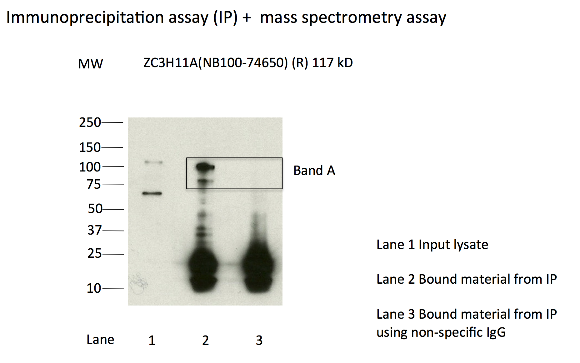
compliant
- Caption
- Immunoprecipitation of ZC3H11A from K562 cells using NB100-74650. Lane 1: input nuclear lysate, Lane 2: material immunoprecipitated with NB100-74650, Lane 3: material immunoprecipitated using control IgG. Bands A was excised from the gel and subject to analysis by mass spectrometry.
- Submitted by
- Kathrina Onate
- Lab
- Michael Snyder, Stanford
- Grant
- U54HG004558
- Download
- IP_K562 Snyder AMN.png
ZC3H11A (Homo sapiens)
MCF-7
Method: immunoprecipitation

compliant
- Caption
- Immunoprecipitation was performed on nuclear extracts from the cell line: MCF-7, using the antibody NB100-74650. The blot shows western blot analysis of input, flowthrough, immunoprecipitate and mock immunoprecipitate using IgG.
- Reviewer comment
- Expected size is around 89 kD but is known to run a bit higher: http://www.novusbio.com/ZC3H11A-Antibody_NB100-74650.html. The image needs lane labels.
- Submitted by
- Denis Salins
- Lab
- Michael Snyder, Stanford
- Grant
- U54HG006996
ZC3H11A (Mus musculus)
Method: immunoprecipitation
not reviewed
- Caption
- Immunoprecipitation of CH12 and MEL nuclear extracts using anti-ZC3H11A antibody (NB100-74650) enriched a single band of the expected molecular weight of Max (~117 kD).
- Submitted by
- Michael Snyder
- Lab
- Michael Snyder, Stanford
- Grant
- RC2HG005602
- Download
- mouse_ZC3H11A_validation_Snyder.pdf
ZC3H11A (Mus musculus)
Method: immunoprecipitation followed by mass spectrometry
not reviewed
- Caption
- This antibody has been validated by IP-Mass Spec human cell lines. Please see the validation document for this antibody in human cell lines for details.
- Submitted by
- Michael Snyder
- Lab
- Michael Snyder, Stanford
- Grant
- RC2HG005602
- Download
- mouse_ZC3H11A_validation_Snyder.pdf
ZC3H11A (Homo sapiens)
A549
Method: immunoprecipitation
exempt from standards
- Caption
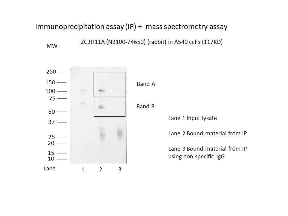
- Immunoprecipitation was performed on nuclear extracts from the cell line A549 using the antibody NB100-74650. Lane 1: input nuclear lysate. Lane 2: material immunoprecipitated with antibody. Lane 3: material immunoprecipitated using control IgG. Marked bands were excised from gel and subjected to analysis by mass spectrometry. Target molecular weight: 117.0.
- Submitter comment
- We showed that TF is detected in both bands by mass-spec.
- Reviewer comment
- Multiple immunoreactive bands seen, but mass-spec secondary characterization demonstrated both detected the factor of interest.
- Submitted by
- Nathaniel Watson
- Lab
- Michael Snyder, Stanford
- Grant
- U54HG006996
- Download
- MS1045_2_ZC3H11A-NB100-74650.JPG
ZC3H11A (Homo sapiens)
GM12878K562HeLa-S3HepG2
Method: immunoblot
compliant
- Caption
- A. Western blots on nuclear lysates from cell lines GM12878 (Lane1), K562 (Lane2), HeLaS3 (Lane3), and HepG2 (Lane4). Expected band size of ZC3H11A is ~89 kD for all cell lines provided.
- Reviewer comment
- Expected size is ~89kD but is also known to run around 117kD and major bands were detected at both sizes.
- Submitted by
- Kathrina Onate
- Lab
- Michael Snyder, Stanford
- Grant
- U54HG004558
- Download
- WB Snyder AMN.png
ZC3H11A (Homo sapiens)
Method: immunoprecipitation followed by mass spectrometry
compliant
- Caption
- IP followed by mass spectrometry. Briefly, protein was immunoprecipitated from A549 nuclear cell lysates using the antibody NB100-74650, and the IP fraction was loaded on a 10% polyacrylamide gel (NuPAGEBis-Tris Gel) and separated with an Invitrogen NuPAGE electrophoresis system. The gel was stained by ColloidialCoomassie G-250 stain, gel fragments corresponding to the bands indicated were excised. Then proteins were trypsinized using the in-gel digestion method. Digested proteins were analyzed on an Orbitrap Elite mass spectrometer (Thermo Scientific) by the nanoLC-ESI-MS/MS technique. Peptides were identified by the SEQUEST algorithm and filtered with a high confidence threshold (Peptide false discovery rate < 1%, 2 unique peptides per protein minimum, mass error < 10 ppm).
- Submitter comment
- None of the proteins ranked above or equal to ZC3H11A have been shown to bind DNA in a sequence-specific manner.
- Reviewer comment
- Both immunoreactive bands were analyzed, as required by the standards.
- Submitted by
- Nathaniel Watson
- Lab
- Michael Snyder, Stanford
- Grant
- U54HG006996
- Download
- ZC3H11A_NB100-74650 final.pdf