ENCAB000AMF
Antibody against Mus musculus USF1, Homo sapiens USF1
Mus musculus
at least one cell type or tissue
awaiting characterization
Homo sapiens
at least one cell type or tissue
awaiting characterization
- Status
- released
- Source (vendor)
- Santa Cruz Biotech
- Product ID
- sc-229
- Lot ID
- A2109
- Characterized targets
- USF1 (Mus musculus), USF1 (Homo sapiens)
- Host
- rabbit
- Clonality
- polyclonal
- External resources
Characterizations
USF1 (Mus musculus)
Method: immunoprecipitation
not reviewed
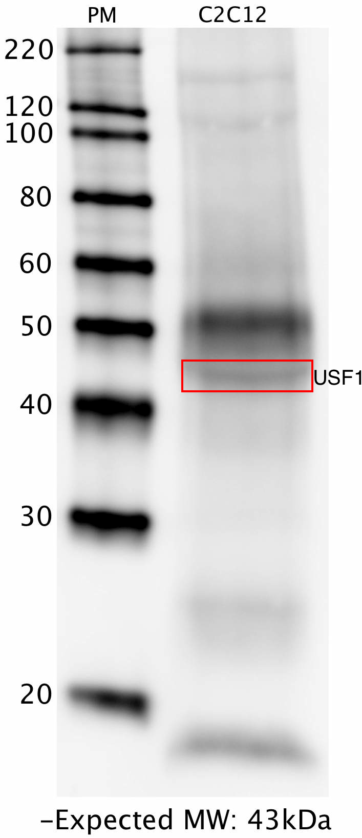
- Caption
- Whole lysate from C2C12 cells was immunoprecipitated with the USF1 (Santa Cruz Biotech, sc-229) antibody. The IP fraction was loaded on a 12% acrylamide gel and separated with a Bio-Rad PROTEAN II xi system. After separation, the samples were transferred to a nitrocellulose membrane with an Invitrogen iBlot system. The membrane was then blotted with primary antibody (same as that used for IP) and then a secondary HRP-conjugated antibody. The resulting bands were visualized using SuperSignal West Femto solution (Thermo Scientific). A band of expected size for the USF1 target (~43 kD) was detected, along with contamination from the heavy chain (~50 kD) and light chain (~25kD) of the primary antibody used for IP.
- Submitted by
- Flo Pauli-Behn
- Lab
- Richard Myers, HAIB
- Grant
- U54HG004576
- Download
- HAIB_USF1_sc-229_mouse_IPwestern.png
USF1 (Homo sapiens)
Method: immunoprecipitation
not reviewed
- Caption
- Western blot protocol: Whole cell lysate was immunoprecipitated using primary antibody, and the IP fraction was loaded on a 12% acrylamide gel and separated with a Bio-Rad PROTEAN II xi system. After separation, the samples were transferred to a nitrocellulose membrane with an Invitrogen iBlot system. Blotting with primary (same as that used for IP) and secondary HRP-conjugated antibodies was performed on an Invitrogen BenchPro 4100 system. Visualization was achieved using SuperSignal West Femto solution (Thermo Scientific). Results: Band of expected size visualized, representing strongest signal in the lane. Figure legend: IP-western with sc-229 in whole cell lysate (WCL) of HeLa, HepG2, K562 and GM12878 ; PM=protein marker. USF1 bands are indicated.
- Submitted by
- Richard Myers
- Lab
- Richard Myers, HAIB
- Grant
- U54HG004576
USF1 (Homo sapiens)
Method: immunoprecipitation followed by mass spectrometry
not reviewed
- Caption
- IP followed by mass spectrometry: Briefly, GM12878 whole cell lysates were immunoprecipitated using primary antibody, and the IP fraction was loaded on a 12% acrylamide gel and separated with a Bio-Rad PROTEAN II xi system. Gel was stained with Coomassie Blue in order to visualize marker bands. Gel fragments corresponding to the bands indicated above in the western blot image were excised and sent to the University of Alabama at Birmingham Cancer Center Mass Spectrometry/Proteomics Shared Facility. There the samples were run on an LTQ XL Linear Ion Trap Mass Spectrometer by LC-ESI-MS/MS. Peptides were identified using SEQUEST tandem mass spectra analysis, with probability based matching at p < 0.05. As per ENCODE data standards, all SEQUEST results are attached (ENCODE_HAIB_USF1_sc229_09122011_MassSpec.pdf), including common contaminants. Target protein is listed as hit 23 in the ~43 kDa band.
- Submitted by
- Richard Myers
- Lab
- Richard Myers, HAIB
- Grant
- U54HG004576