ENCAB000AKR
Antibody against Homo sapiens SIN3A, Mus musculus SIN3A
Homo sapiens
MCF-7, GM12878, K562, HeLa-S3, HepG2
characterized to standards
Homo sapiens
A549
characterized to standards with exemption
Homo sapiens
any cell type or tissue
partially characterized
Mus musculus
any cell type or tissue
partially characterized
- Status
- released
- Source (vendor)
- Novus
- Product ID
- NB600-1263
- Lot ID
- LB140235
- Characterized targets
- SIN3A (Homo sapiens), SIN3A (Mus musculus)
- Host
- rabbit
- Clonality
- polyclonal
- Purification
- other
- Antigen description
- Immunogen: Synthetic peptide: MKRRLDDQESDVYAAQQRR, corresponding to amino acids 1-19 of Mouse mSin3A.
- Antigen sequence
- MKRRLDDQESDVYAAQQRR
- External resources
Characterizations
SIN3A (Homo sapiens)
A549
Method: immunoprecipitation
exempt from standards
- Caption
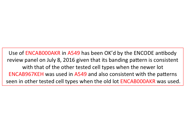
- Use of ENCAB000AKR in A549 has been OK’d by the ENCODE antibody review panel on July 8, 2016 given that its banding pattern is consistent with that of the other tested cell types when the newer lot ENCAB967KEH was used in A549 and also consistent with the patterns seen in other tested cell types when the old lot ENCAB000AKR was used.
- Submitter comment
- We have run out of this antibody lot and cannot do a primary in A549. Instead, we have done primaries using a new lot in A549 and other cell types (ENCAB967KEH).
- Reviewer comment
- This exemption request was forwarded to the ENCODE antibody review panel and was granted on July 8, 2016.
- Submitted by
- Esther Chan
- Lab
- Michael Snyder, Stanford
- Grant
- U54HG006996
- Download
- ENCAB000AKR_A549_primary.png
SIN3A (Homo sapiens)
K562
Method: immunoprecipitation
compliant
- Caption
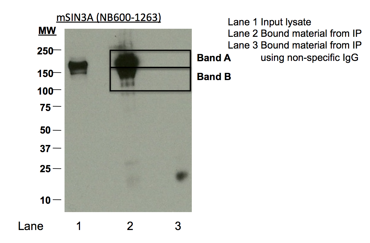
- Immunoprecipitation of SIN3A from K562 cells using NB600-1263. Lane 1: input nuclear lysate. Lane 2: immunoprecipitated material. lane 3: material immunoprecipitated with control IgG. Bands A and B were excised from the gel and subject to analysis by mass spectrometry.
- Submitted by
- Kathrina Onate
- Lab
- Michael Snyder, Stanford
- Grant
- U54HG004558
- Download
- ENCAB000AKR_K562_IP.png
SIN3A (Homo sapiens)
Method: immunoprecipitation
not reviewed
- Caption
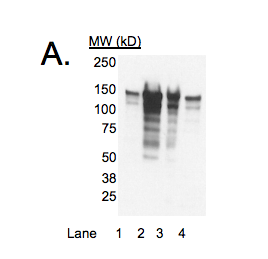
- In Western blots, a major band of the expected size of SIN3A (~145kD) is observed in cell lines GM12878, K562, HeLa S3, and HepG2 (Panel A). This band is efficiently and specifically immunoprecipitated from K562 nuclear lysates (arrow, Panel B). We note that additional bands are present, particularly in cell line K562 and HepG2, but these are significantly less abundant (see Panel B) and can be identified as SIN3A by mass spectrometry (see validation #2). Therefore, NB600-1263 meets this criterion for validation.
- Submitted by
- Michael Snyder
- Lab
- Michael Snyder, Stanford
- Grant
- U54HG004558
SIN3A (Homo sapiens)
Method: ChIP-seq comparison
not reviewed
- Submitted by
- Michael Snyder
- Lab
- Michael Snyder, Stanford
- Grant
- U54HG004558
SIN3A (Homo sapiens)
K562
Method: immunoprecipitation
compliant
- Caption
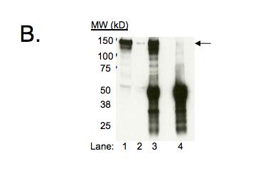
- B. Immunoprecipitation of SIN3A from K562 cells using NB600-1263. Lane 1: input nuclear lysate, Lane 2: unbound material from immunoprecipitation with NB600-1263, Lane 3: material immunoprecipitated with NB600-1263, Lane 4: material immunoprecipitated using control IgG. Arrow indicates specifically immunoprecipitated band.
- Submitted by
- Kathrina Onate
- Lab
- Michael Snyder, Stanford
- Grant
- U54HG004558
- Download
- IP Snyder AKR.png
SIN3A (Homo sapiens)
MCF-7
Method: immunoprecipitation
compliant
- Caption
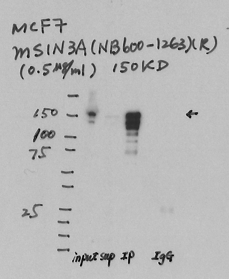
- Immunoprecipitation was performed on nuclear extracts from the cell line: MCF-7, using the antibody NB600-1263. The blot shows western blot analysis of input, flowthrough, immunoprecipitate and mock immunoprecipitate using IgG.
- Reviewer comment
- Lanes not labeled and hard to interpret.
- Submitted by
- Denis Salins
- Lab
- Michael Snyder, Stanford
- Grant
- U54HG006996
SIN3A (Mus musculus)
Method: immunoprecipitation
not reviewed
- Submitted by
- Michael Snyder
- Lab
- Michael Snyder, Stanford
- Grant
- RC2HG005602
SIN3A (Homo sapiens)
Method: immunoprecipitation followed by mass spectrometry
compliant
- Caption
- IP followed by mass spectrometry: Briefly, protein was immunoprecipitated from K562 whole cell lysates using NB600-1263, and the IP fraction was loaded on a 10% polyacrylamide gel (NuPAGE Bis-Tris Gel) and separated with an Invitrogen NuPAGE electrophoresis system. The gel was silver-stained, gel fragments corresponding to the bands indicated were excised and destained using the SilverSNAP Stain for Mass Spectrometry (Pierce). Then proteins were treated with trypsin using the in-gel digestion method. Digested proteins were analyzed on a LTQ-Orbitrap (Thermo Scientific) b the nanoLC-ESI-MS/MS technique. Peptides were identified by the SEQUEST algorithm and filtered with a high confidence threshold (protein false discovery rate <1%, 2 peptides per protein minimum).
- Submitted by
- Kathrina Onate
- Lab
- Michael Snyder, Stanford
- Grant
- U54HG004558
- Download
- Sin3A_final_AKR Sheet1.pdf
SIN3A (Homo sapiens)
GM12878K562HeLa-S3HepG2
Method: immunoblot
compliant
- Caption
- A. Western blot using antibody NB600-1263. Lanes contain nuclear lysates from GM12878 cells (Lane 1), K562 cells (Lane 2), HeLa S3 cells (Lane 3), and HepG2 cells (Lane 4). Expected band size of SIN3A for all cell lines is ~145 kD.
- Reviewer comment
- Lane 2 (K562) has multiple bands with the major band not at least 50% of the total signal.
- Submitted by
- Kathrina Onate
- Lab
- Michael Snyder, Stanford
- Grant
- U54HG004558
- Download
- WB Snyder AKR.png