ENCAB681JHP
Antibody against Homo sapiens MITF
Homo sapiens
K562
partially characterized
Homo sapiens
at least one cell type or tissue
not pursued
- Status
- released
- Source (vendor)
- Active Motif
- Product ID
- 39789
- Lot ID
- 11313002
- Characterized targets
- MITF (Homo sapiens)
- Host
- mouse
- Clonality
- monoclonal
- Purification
- Protein G
- Aliases
- michael-snyder:AS-819
- External resources
Characterizations
MITF (Homo sapiens)
K562
Method: immunoprecipitation
not compliant
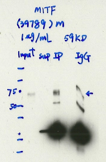
- Caption
- Immunoprecipitation was performed on nuclear extracts from the cell line: K562, using the antibody 39789. The blot shows western blot analysis of input, flowthrough, immunoprecipitate and mock immunoprecipitate using IgG.
- Reviewer comment
- IgG appears not different from the IP
- Submitted by
- Denis Salins
- Lab
- Michael Snyder, Stanford
- Grant
- U54HG006996
- Download
- Expt 825_1-MITF.jpg
MITF (Homo sapiens)
K562
Method: immunoprecipitation

not compliant
- Caption
- Immunoprecipitation was performed on nuclear extracts from the cell line: K562, using the antibody 39789. The blot shows western blot analysis of input, flowthrough, immunoprecipitate and mock immunoprecipitate using IgG.Molecular Weight: 58.795
- Reviewer comment
- Band is not clearly absent from the IgG control lane
- Submitted by
- Nathaniel Watson
- Lab
- Michael Snyder, Stanford
- Grant
- U54HG006996
- Download
- K562-MITF.jpg
MITF (Homo sapiens)
K562
Method: immunoprecipitation
compliant
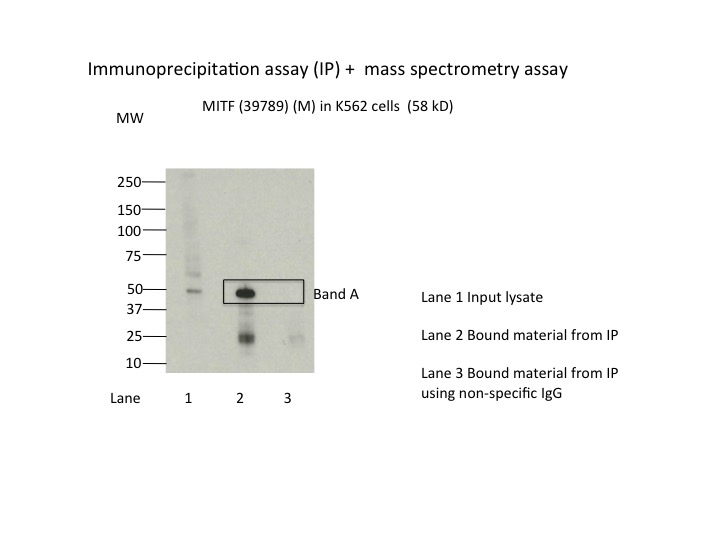
- Caption
- Immunoprecipitation was performed on nuclear extracts from the cell line K562 using the antibody 39789. Lane 1: input nuclear lysate. Lane 2: material immunoprecipitated with antibody. Lane 3: material immunoprecipitated using control IgG. Marked bands were excised from gel and subjected to analysis by mass spectrometry. Target molecular weight: 58.795.
- Submitted by
- Nathaniel Watson
- Lab
- Michael Snyder, Stanford
- Grant
- U54HG006996
- Download
- MITF.jpg
MITF (Homo sapiens)
Method: immunoprecipitation
not submitted for review by lab
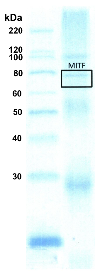
- Caption
- K562 whole cell lysate was immunoprecipitated using the primary antibody (Active Motif; 39789). The IP fraction was loaded on a 12% Bio-Rad TGX gel and separated with the Bio-Rad Tetra Cell system. A gel fragment (rectangle outline) corresponding to the band indicated on the Coomassie Blue stained gel image was excised and sent to the University of Alabama at Birmingham Cancer Center Mass Spectrometry/Proteomics Shared Facility.
- Submitter comment
- This image is to show which bands are used for the mass spec
- Submitted by
- Mark Mackiewicz
- Lab
- Richard Myers, HAIB
- Grant
- U54HG006998
- Download
- MITF_IP_MS_WB_1.png