ENCAB000AMQ
Alternate accession: ENCAB548JNC
Antibody against Homo sapiens ZMIZ1, Mus musculus ZMIZ1
Homo sapiens
K562
characterized to standards
Homo sapiens
any cell type or tissue, HEK293T
partially characterized
Mus musculus
any cell type or tissue
partially characterized
- Status
- released
- Source (vendor)
- Abcam
- Product ID
- ab65767
- Lot ID
- GR30927-1
- Characterized targets
- ZMIZ1 (Homo sapiens), ZMIZ1 (Mus musculus)
- Host
- rabbit
- Clonality
- polyclonal
- Purification
- affinity
- Antigen description
- Raised against synthetic peptide conjugated to KLH derived from within residues 200 - 300 of Human Zinc finger MIZ domain-containing protein 1.
- Aliases
- michael-snyder:AS-1329
- External resources
Characterizations
ZMIZ1 (Homo sapiens)
HEK293T
Method: immunoprecipitation
not submitted for review by lab
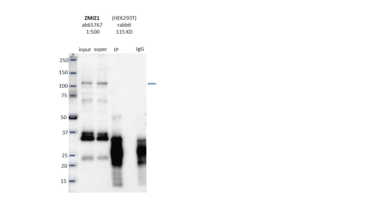
- Caption
- Immunoprecipitation was performed on nuclear extracts from the cell line: HEK293T using the antibody ab65767_GR30927-1. The image shows western blot analysis of input, flowthrough, immunoprecipitate, and mock immunoprecipitate using IgG. Target molecular weight: 115.
- Submitted by
- Nathaniel Watson
- Lab
- Michael Snyder, Stanford
- Grant
- U54HG006996
- Download
- Expt1038_12-ZMIZ1-ab65767.JPG
ZMIZ1 (Homo sapiens)
Method: immunoprecipitation followed by mass spectrometry
not reviewed
- Caption
- Immunoprecipitation of ZMIZ1 from K562 cells using ab65767. Lane 1: input nuclear lysate, Lane 2: material immunoprecipitated with ab65767, Lane 3: material immunoprecipitated using control IgG. Bands A was excised from the gel and subject to analysis by mass spectrometry. This antibody was raised against an immunogen that is predicted to cross react with both isoform 1 (115 kDa) and isoform 2 (107 kDa) of ZMIZ1. The bands we observe at ~115 kDa and ~105 kDa could possibly correspond to teh two isoforms of ZMIZ1. IP followed by mass spectrometry: Briefly, protein was immunoprecipitated from K562 whole cell lysates using ab65767, and the IP fraction was loaded on a 10% polyacrylamide gel (NuPAGE Bis-Tris Gel) and separated with an Invitrogen NuPAGE electrophoresis system. The gel was silver-stained, gel fragments corresponding to the bands indicated were excised and destained using the SilverSNAP Stain for Mass Spectrometry (Pierce). Then proteins were trypsinized using the in-gel digestion method. Digested proteins were analyzed on an LTQ-Orbitrap (Thermo Scientific) by the nanoLC-ESI-MS/MS technique. Peptides were identified by the SEQUEST algorithm and filtered with a high confidence threshold (Protein false discovery rate < 1%, 2 peptides per protein minimum). We report 22 different proteins identified in band A, of which 9 were detected in the control IgG IP too, indicating a non specific enrichment of these proteons during immunoprecipitation. 3 out of the top 5 hits were enriched in both the ab65767 as well as control IgG IP. Of the specifically immunoprecipitated proteins, ZMIZ1 is the most abundant protein. Based on these observations, this band is likely due to the presence of immunoprecipitated ZMIZ1 and ab65767 meets the ENCODE standard for validation by this criterion.
- Submitted by
- Michael Snyder
- Lab
- Michael Snyder, Stanford
- Grant
- U54HG004558
ZMIZ1 (Homo sapiens)
Method: immunoprecipitation
not reviewed
- Caption
- A. Western blots on nuclear lysates from cell lines GM12878 (Lane1), K562 (Lane2), HeLaS3 (Lane3), and HepG2 (Lane4). B. Immunoprecipitation was performed on nuclear lysates from K562 cells using antibody ab65767 against ZMIZ1. Lane1: Nuclear lysate. Lane 2: Unbound material from immunoprecipitation with ab65767. Lane 3: Bound material from immunoprecipitation with ab65767. Lane 4: Bound material from control immunoprecipitation with rabbit IgG. Arrow indicates band of expected size (115kD) that is enriched in the specifically immunoprecipitated fraction. Smaller bands could be possibly degradation products of ZMIZ1 protein. Band indicated by * in K562 immunoprecipitate is IgG light chains. A band of ~115 kD is detected by Western blotting with ab65767 in multiple human cell lines. Immunoprecipitation from K562 nuclear lysate enriches a protein of ~115KD. Based on these observations, this antibody meets this ENCODE criterion.
- Submitted by
- Michael Snyder
- Lab
- Michael Snyder, Stanford
- Grant
- U54HG004558
ZMIZ1 (Homo sapiens)
K562
Method: immunoprecipitation

compliant
- Caption
- Immunoprecipitation of ZMIZ1 from K562 cells using ab65767. Lane 1: input nuclear lysate, Lane 2: material immunoprecipitated with ab65767, Lane 3: material immunoprecipitated using control IgG. Bands A was excised from the gel and subject to analysis by mass spectrometry. This antibody was raised against an immunogen that is predicted to cross react with both isoform 1 (115 kDa) and isoform 2 (107 kDa) of ZMIZ1. The bands we observe at ~115 kDa and ~105 kDa could possibly correspond to the two isoforms of ZMIZ1.
- Submitted by
- Kathrina Onate
- Lab
- Michael Snyder, Stanford
- Grant
- U54HG004558
- Download
- IP-MS_ZMIZ1:K562 Snyder AMQ.png
ZMIZ1 (Mus musculus)
Method: immunoprecipitation followed by mass spectrometry
not reviewed
- Caption
- This antibody has been validated by IP-Mass Spec in multiple human cell lines. Please see the validation document for this antibody in human cell lines for details.
- Submitted by
- Michael Snyder
- Lab
- Michael Snyder, Stanford
- Grant
- RC2HG005602
- Download
- mouse_ZMIZ1_validation_Snyder.pdf
ZMIZ1 (Mus musculus)
Method: immunoprecipitation
not reviewed
- Caption
- This antibody has been validated by IP-Mass Spec in multiple human cell lines. Please see the validation document for this antibody in human cell lines for details.
- Submitted by
- Michael Snyder
- Lab
- Michael Snyder, Stanford
- Grant
- RC2HG005602
- Download
- mouse_ZMIZ1_validation_Snyder.pdf
ZMIZ1 (Homo sapiens)
Method: immunoprecipitation followed by mass spectrometry
compliant
- Caption
- IP followed by mass spectrometry: Briefly, protein was immunoprecipitated from K562 whole cell lysates using ab65767, and the IP fraction was loaded on a 10% polyacrylamide gel (NuPAGE Bis-Tris Gel) and separated with an Invitrogen NuPAGE electrophoresis system. The gel was silver-stained, gel fragments corresponding to the bands indicated were excised and destained using the SilverSNAP Stain for Mass Spectrometry (Pierce). Then proteins were trypsinized using the in-gel digestion method. Digested proteins were analyzed on an LTQ-Orbitrap (Thermo Scientific) by the nanoLC-ESI-MS/MS technique. Peptides were identified by the SEQUEST algorithm and filtered with a high confidence threshold (Protein false discovery rate < 1%, 2 peptides per protein minimum). We report 22 different proteins identified in band A, of which 9 were detected in the control IgG IP too, indicating a non specific enrichment of these proteons during immunoprecipitation. 3 out of the top 5 hits were enriched in both the ab65767 as well as control IgG IP. Of the specifically immunoprecipitated proteins, ZMIZ1 is the most abundant protein.
- Submitted by
- Kathrina Onate
- Lab
- Michael Snyder, Stanford
- Grant
- U54HG004558
- Download
- ZMIZ1_final Sheet2 (3).pdf