ENCAB210NHK
Antibody against Mus musculus CTCF
Mus musculus
at least one cell type or tissue
awaiting characterization
- Status
- released
- Source (vendor)
- Victor Lobanenkov
- Product ID
- CTCF
- Lot ID
- unknown
- Characterized targets
- CTCF (Mus musculus)
- Host
- mouse
- Clonality
- monoclonal
- Antigen description
- Mouse monoclonal antibodies were raised against human recombinant CTCF expressed in Pichia pastoris. Protein for immunization was prepared as described (1). Briefly, recombinant yeasts were induced with methanol and then were washed and frozen at ––80oC. Cells were homogenized in a homogenizing buffer (40mM HEPES, pH 7.6, 2mM MgCl2, 0.1mM EDTA, 100uM ZnSO4, 5% glycerol, 100mM KCl, 5mM DTT and 1M urea) with subsequent centrifugations at 16000g for 20 min and 35000g for 90 min. Supernatants were filtered and were subjected to three steps of chromatography on SP-Sepharose, Ni2+-Sepharose and heparin-Sepharose. For immunization CTCF was further purified by SDS-PAGE and the band was cut from the gel and was injected into mice directly. For the screening purposes we used protein after chromatography. Total of 9 clones were isolated and antibodies were checked in two assays –– immunoblotting and EMSA supershift. The results of both assays were presented on the Supplementary Fig.1 panels A and B. Total cell extracts from Jurkat cells were loaded in duplicates and stained with antibodies (total ascites) in dilution 1:100. All 9 antibodies showed specific staining of 140kDa band corresponding to CTCF, while negative control, non-specific rabbit IgG antibody, didn’t detect specific CTCF band. Some minor bands did show up with different antibodies, which might be staining of degradation products. All 9 antibodies also alter EMSA CTCF band suggesting that all of them can recognize native protein bound to DNA. To increase the efficiency of ChIP assay we used a mixture of all 9 CTCF antibodies to perform ChIPs with CTCF. This mixture shows far superior results in terms of reproducibility then all polyclonal antibodies tested so far (Upstate Biotechnologies and Abcam). More information on the antibody can be found in publication PMID:15731119.
- External resources
Characterizations
CTCF (Mus musculus)
Method: immunoblot
not reviewed
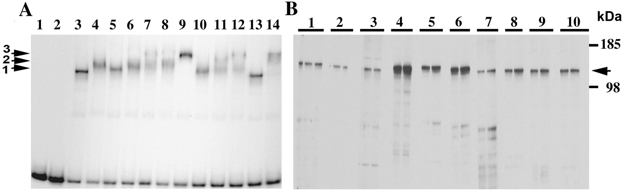
- Caption
- (A) EMSA with newly developed CTCF monoclonal antibodies. Lane 1- free probe, lane 2- ivt luciferase negative control, lane 3- ivt CTCF, lane 4- ivt CTCF + Ab2.24.3.10.1.4, lane 5- ivt CTCF + Ab2.22.1.2.2.3, lane 6- ivt CTCF + Ab2.27.2.5.3.2.5, lane 7- ivt CTCF + Ab2.17.1.1.4, lane 8- ivt CTCF + Ab2.12.3.6.2.11.11, lane 9- ivt CTCF + Ab1.1.2.29.9.11, lane 10- ivt CTCF + Ab1.3.2.5.5.2, lane 11- ivt CTCF + Ab2.30.3.19.6.1, lane 12- ivt CTCF + Ab1.2.15.4, lane 13- ivt CTCF + Ab negative control (non-specific rabbit IgG), lane 13- ivt CTCF + Ab positive control (anti-CTCF MABs, purchased from BD Transduction Laboratories with catalog number 612149). Human mutant Xist C(-43)G promoter fragment was used as a probe for EMSA. Arrow 1 shows specific CTCF shift in the lanes 1 and 13, without CTCF antibodies and with non-specific rabbit antibodies, respectively. Arrows 2 and 3 show super-shifts in the lanes with CTCF antibodies, thereby confirming specific CTCF binding to the C(-43)G mutant XIST promoter probe. (B) Immunoblot analysis of nine CTCF monoclonal antibodies. Equal amounts of Jurkat cell lysates were loaded in each well. After transfer to nitrocellulose two wells were cut and probed with each antibody. 1- Ab2.24.3.10.1.4, 2- Ab2.22.1.2.2.3, 3- Ab2.27.2.5.3.2.5, 4- Ab2.17.1.1.4, 5- Ab2.12.3.6.2.11.11, 6- Ab1.1.2.29.9.11, 7- Ab1.3.2.5.5.2, 8- Ab2.30.3.19.6.1, 9- Ab1.2.15.4, 10- Ab positive control (BD anti-CTCF MABs). The black arrow shows the major endogenous CTCF protein with molecular mass about 140 kDa. The rainbow molecular mass protein markers are indicated by numbers 185 and 98 kDa on the right of panel B.
- Submitted by
- Yin Shen
- Lab
- Bing Ren, UCSD
- Grant
- R01HG003991