ENCAB000AIH
Alternate accession: ENCAB827NOG
Antibody against Homo sapiens KDM1A
Homo sapiens
K562, HepG2, GM12878, endothelial cell of umbilical vein, A549, H1
characterized to standards
- Status
- released
- Source (vendor)
- Bethyl Labs
- Product ID
- A300-215A
- Lot ID
- 1
- Characterized targets
- KDM1A (Homo sapiens)
- Host
- rabbit
- Clonality
- polyclonal
- Purification
- affinity
- Aliases
- bradley-bernstein:PchAb 158
- External resources
Characterizations
KDM1A (Homo sapiens)
K562
Method: immunoblot
compliant
- Submitted by
- Bradley Bernstein
- Lab
- Bradley Bernstein, Broad
- Grant
- U54HG004570
- Download
- human_LSD1_validation_Bernstein.pdf
KDM1A (Homo sapiens)
Method: ChIP-seq comparison
not reviewed
- Submitted by
- Bradley Bernstein
- Lab
- Bradley Bernstein, Broad
- Grant
- U54HG004570
- Download
- human_LSD1_validation_Bernstein.pdf
KDM1A (Homo sapiens)
Method: ChIP-seq comparison
compliant
- Caption
- This validation relies on the use of antibodies to different members of a known complex, and the demonstration that highly similar patterns of enrichment are obtained with each antibody. The second track shown used an antibody to CHD4 (PchAb 1222), another member of the NuRD complex. Overall correlation score: KDM1A (also known as LSD1) vs CHD4: 0.8585
- Submitted by
- Kathrina Onate
- Lab
- Bradley Bernstein, Broad
- Grant
- U54HG006991
KDM1A (Homo sapiens)
Method: ChIP-seq comparison
not compliant
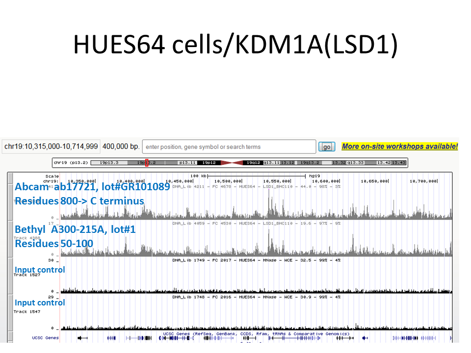
- Caption
- ChIP-seq was performed twice using HUES64 cells, using two different antibodies that were raised against different regions of the same protein (KDM1A), and highly similar results were obtained. This is suggestive that the enrichment obtained using ChIP is due to a specific interaction between the antibody and the intended cellular protein.
- Reviewer comment
- Failed due to needing more information (needs to be compared to an eligible antibody, needs IDR details, needs ENCAB and ENCSR info about antibody used to compare)
- Submitted by
- Noam Shoresh
- Lab
- Bradley Bernstein, Broad
- Grant
- U54HG006991
KDM1A (Homo sapiens)
A549H1
Method: immunoblot
compliant
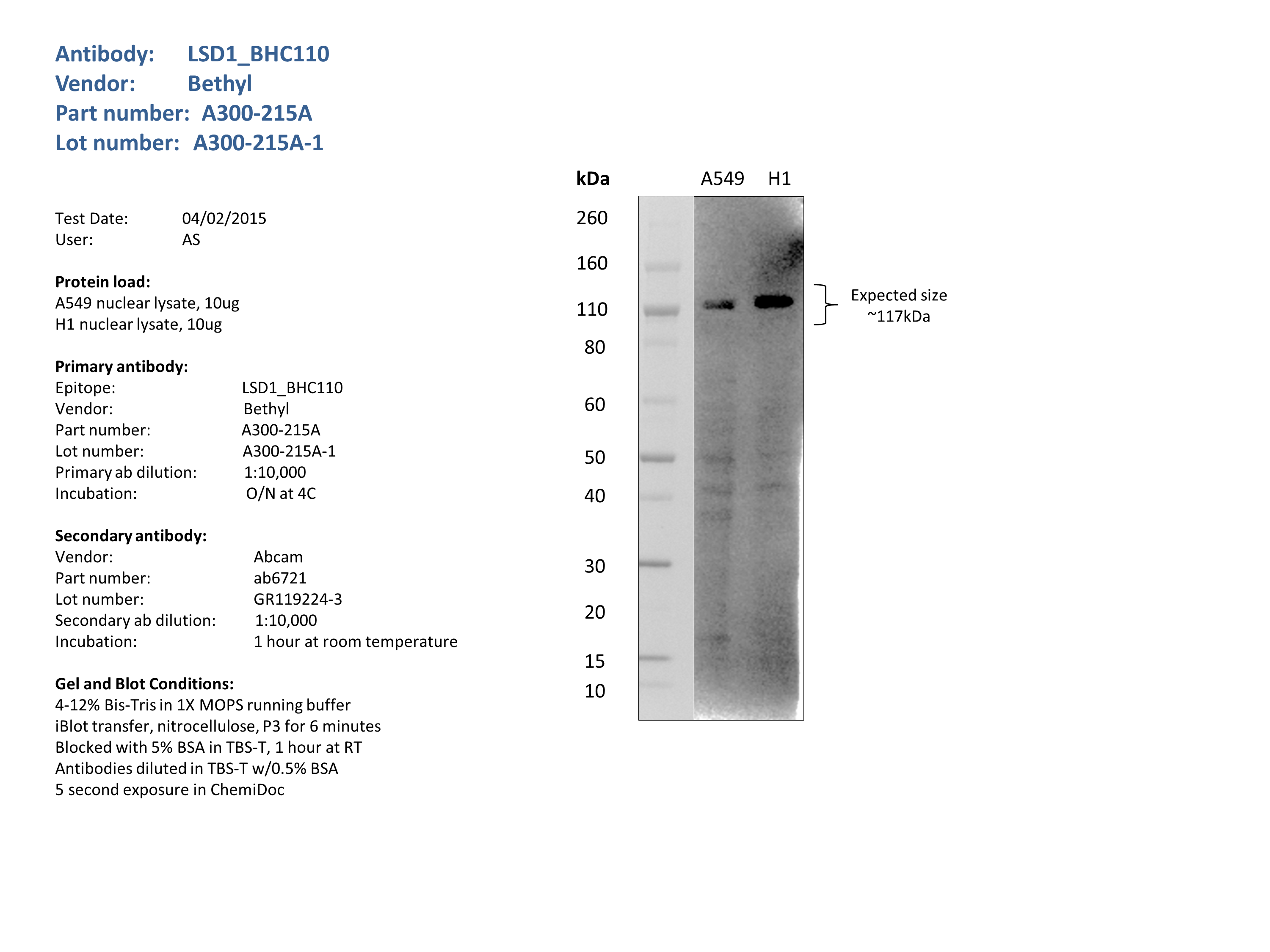
- Caption
- Nuclear lysates from A549 (10ug) and H1 (10ug) were loaded into a 4-12% Bis-Tris gel in 1X MOPS running buffer. After separation, the samples were transferred to a nitrocellulose membrane using iblot. Membrane was blocked for an hour in room temperature, with 5% BSA in TBS-T and blotted with primary antibody in the appropriate concentration over night at 4c. Membrane was washed and blotted with secondary HRP-conjugated antibody. Detection was made with Optiblot ECL Detect Kit (ab133406) for 2 min. Bands were detected at the expected size.
- Submitted by
- Noam Shoresh
- Lab
- Bradley Bernstein, Broad
- Grant
- U54HG006991
KDM1A (Homo sapiens)
K562HepG2GM12878endothelial cell of umbilical vein
Method: immunoblot
compliant
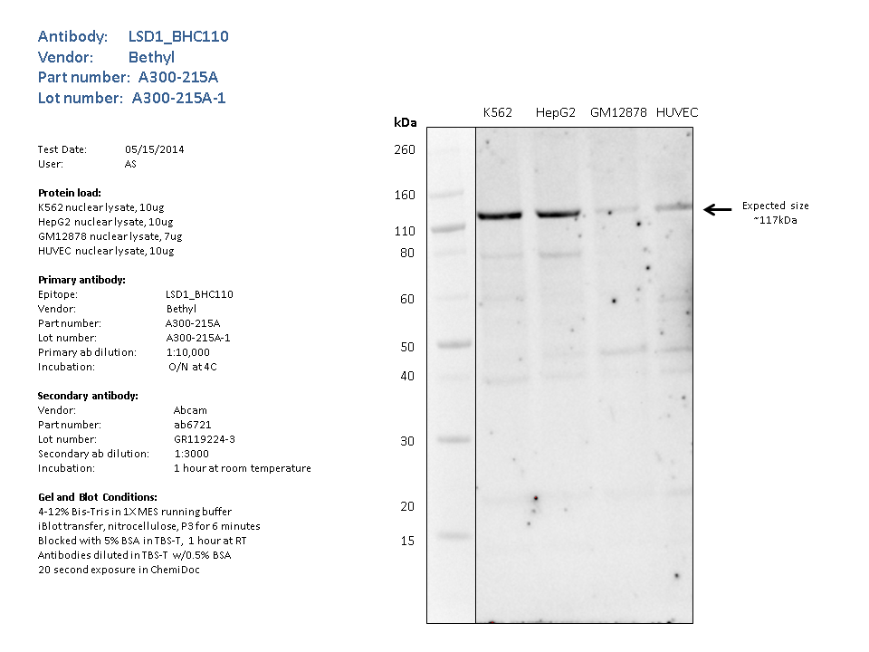
- Caption
- Nuclear lysates from K562 (10ug), HepG2 (10ug), GM12878 (7ug), HUVEC (10ug), were loaded into a 4-12% Bis-Tris gel in 1X MES running buffer. After separation, the samples were transferred to a nitrocellulose membrane using the iblot system. Membrane was blocked for an hour in room temperature, with 5% BSA in TBS-T and blotted with primary antibody in the appropriate concentration over night at 4c. Membrane was washed and blotted with secondary HRP-conjugated antibody. Detection was made with Optiblot ECL Detect Kit (ab133406) for 2 min.the strongest band was detected around the expected size (~117kDa).
- Submitted by
- Noam Shoresh
- Lab
- Bradley Bernstein, Broad
- Grant
- U54HG006991