ENCAB000AHE
Antibody against Homo sapiens GTF2F1
Homo sapiens
K562, HeLa-S3, HepG2, MCF-7
characterized to standards
Homo sapiens
any cell type or tissue
partially characterized
Homo sapiens
GM12878
not characterized to standards
- Status
- released
- Source (vendor)
- Abcam
- Product ID
- ab28179
- Lot ID
- GR60531
- Characterized targets
- GTF2F1 (Homo sapiens)
- Host
- rabbit
- Clonality
- polyclonal
- Antigen description
- Recombinant full length protein - RAP 74 subunit of TF11F.
- External resources
Characterizations
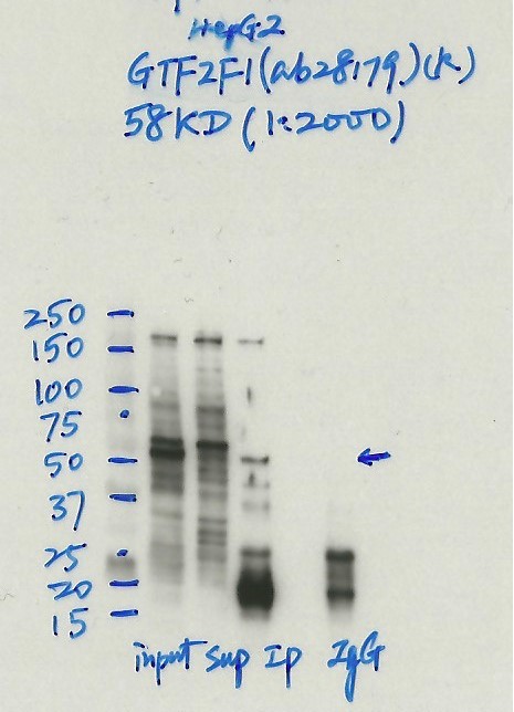
GTF2F1 (Homo sapiens)
HepG2
Method: immunoprecipitation
compliant
- Caption
- Immunoprecipitation was performed on nuclear extracts from the cell line: HepG2, using the antibody ab28179. The blot shows western blot analysis of input, flowthrough, immunoprecipitate and mock immunoprecipitate using IgG.Molecular Weight: 58 kDa.
- Submitted by
- Nathaniel Watson
- Lab
- Michael Snyder, Stanford
- Grant
- U54HG006996
- Download
- #1132 HepG2 GTF2F1(ab28179) (2).jpg
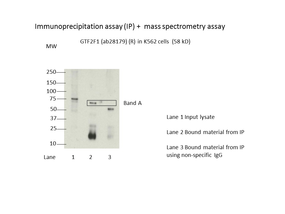
GTF2F1 (Homo sapiens)
K562
Method: immunoprecipitation
compliant
- Caption
- Immunoprecipitation was performed on nuclear extracts from the cell line K562 using the antibody ab28179. Lane 1: input nuclear lysate. Lane 2: material immunoprecipitated with antibody. Lane 3: material immunoprecipitated using control IgG. Marked bands were excised from gel and subjected to analysis by mass spectrometry. Target molecular weight: 58.
- Submitted by
- Nathaniel Watson
- Lab
- Michael Snyder, Stanford
- Grant
- U54HG006996
- Download
- 1009_GTF2F1.jpg
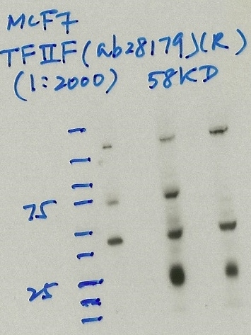
GTF2F1 (Homo sapiens)
MCF-7
Method: immunoprecipitation
compliant
- Caption
- Immunoprecipitation was performed on nuclear extracts from the cell line: MCF-7, using the antibody ab28179. The blot shows western blot analysis of input, flowthrough, immunoprecipitate and mock immunoprecipitate using IgG.
- Reviewer comment
- The expected size on the image and the target is wrong. This antibody is supposed to target the ~70kD subunit of the TFIIF heterodimer.
- Submitted by
- Denis Salins
- Lab
- Michael Snyder, Stanford
- Grant
- U54HG006996
- Download
- 811 3_ TFIIF.jpg
GTF2F1 (Homo sapiens)
Method: immunoprecipitation
not submitted for review by lab
- Caption
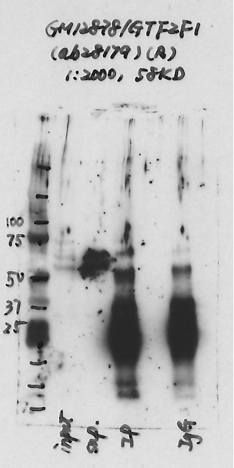
- Immunoprecipitation was performed on nuclear extracts from the cell line: GM12878, using the antibody ab28179. The blot shows western blot analysis of input, flowthrough, immunoprecipitate and mock immunoprecipitate using IgG.Molecular Weight: None
- Submitted by
- Nathaniel Watson
- Lab
- Michael Snyder, Stanford
- Grant
- U54HG006996
- Download
- GM12878-GTF2F1.jpg
GTF2F1 (Homo sapiens)
Method: immunoprecipitation followed by mass spectrometry
compliant
- Caption
- IP followed by mass spectrometry. Briefly, protein was immunoprecipitated from K562 nuclear cell lysates using the antibody ab28179, and the IP fraction was loaded on a 10% polyacrylamide gel (NuPAGEBis-Tris Gel) and separated with an Invitrogen NuPAGE electrophoresis system. The gel was stained by ColloidialCoomassie G-250 stain, gel fragments corresponding to the bands indicated were excised. Then proteins were trypsinized using the in-gel digestion method. Digested proteins were analyzed on an Orbitrap Elite mass spectrometer (Thermo Scientific) by the nanoLC-ESI-MS/MS technique. Peptides were identified by the SEQUEST algorithm and filtered with a high confidence threshold (Peptide false discovery rate < 1%, 2 unique peptides per protein minimum, mass error < 10 ppm).
- Submitter comment
- Biogrid: GTF2F1 has interaction with TAT (4 publication), MATR3 also has interaction with TAT (1 publication).http://thebiogrid.org/109217/summary/homo-sapiens/gtf2f1.html, http://thebiogrid.org/115126/summary/homo-sapiens/matr3.html. MATR3 is the the same gel slice with GTF2F1, but TAT is not. Biogrid: GTF2F1 has interaction with PARP1 (2 publication), XRCC5 also has interaction with PARP1 (9 publication).http://thebiogrid.org/109217/summary/homo-sapiens/gtf2f1.html; http://thebiogrid.org/113353/summary/homo-sapiens/xrcc5.html. XRCC5 is the same gel slice with GTF2F1, but PARP1 is not.
- Submitted by
- Nathaniel Watson
- Lab
- Michael Snyder, Stanford
- Grant
- U54HG006996
- Download
- GTF2F1_ab28179 final.pdf
GTF2F1 (Homo sapiens)
Method: immunoblot
not reviewed
- Submitted by
- Michael Snyder
- Lab
- Michael Snyder, Stanford
- Grant
- U54HG004558
GTF2F1 (Homo sapiens)
Method: immunoprecipitation
not reviewed
- Caption
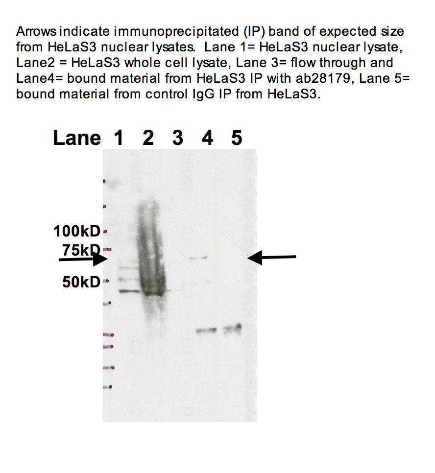
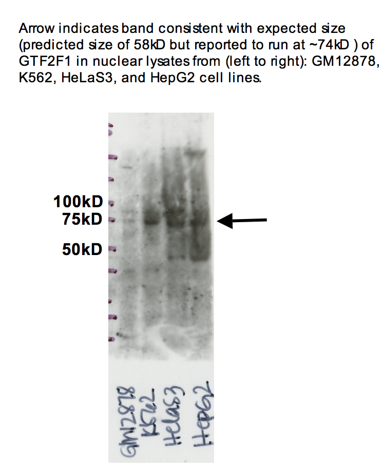
- Arrow indicates band consistent with expected size (predicted size of 58kD but reported to run at ~74kD ) of GTF2F1 in nuclear lysates from (left to right): GM12878, K562, HeLaS3, and HepG2 cell lines. 100kD 75kD 50kd Arrows indicate immunoprecipitated (IP) band of expected size from HeLaS3 nuclear lysates. Lane 1= HeLaS3 nuclear lysate, Lane2 = HeLaS3 whole cell lysate, Lane 3= flow through and Lane4= bound material from HeLaS3 IP with ab28179, Lane 5= bound material from control IgG IP from HeLaS3.
- Submitted by
- Michael Snyder
- Lab
- Michael Snyder, Stanford
- Grant
- U54HG004558
GTF2F1 (Homo sapiens)
HeLa-S3
Method: immunoprecipitation
compliant
- Caption
- Arrows indicate immunoprecipitated (IP) band of expected size from HeLaS3 nuclear lysates. Lane 1= HeLaS3 nuclear lysate, Lane2 = HeLaS3 whole cell lysate, Lane 3= flow through and Lane4= bound material from HeLaS3 IP with ab28179, Lane 5= bound material from control IgG IP from HeLaS3.
- Reviewer comment
- HepG2 compliant
- Submitted by
- Kathrina Onate
- Lab
- Michael Snyder, Stanford
- Grant
- U54HG004558
- Download
- IP Snyder AHE.png
GTF2F1 (Homo sapiens)
GM12878K562HeLa-S3HepG2
Method: immunoblot
compliant
- Caption
- Arrow indicates band consistent with expected size (predicted size of 58kD but reported to run at ~74kD ) of GTF2F1 in nuclear lysates from (left to right): GM12878, K562, HeLaS3, and HepG2 cell lines. 100kD 75kD 50kd Arrows indicate immunoprecipitated (IP) band of expected size from HeLaS3 nuclear lysates. Lane 1= HeLaS3 nuclear lysate, Lane2 = HeLaS3 whole cell lysate, Lane 3= flow through and Lane4= bound material from HeLaS3 IP with ab28179, Lane 5= bound material from control IgG IP from HeLaS3.
- Reviewer comment
- 1, 3, 4 not compliant; 2 compliant
- Submitted by
- Kathrina Onate
- Lab
- Michael Snyder, Stanford
- Grant
- U54HG004558
- Download
- WB Snyder AHE.png