ENCBS458AAA / cell line
Summary
- Status
- released
- Term name
- K562
- Term ID
- EFO:0002067
- Summary
- Homo sapiens K562 cell line stably expressing C-terminal eGFP-tagged ATF3
- Description
- K562 cell line stably expressing a C-terminal LAP-tag containing eGFP, fused to ATF3-L
- Culture start date
- 2014-10-29
- Culture harvest date
- 2014-11-21
- Passage number
- 6
Attribution
ENCODE3 project
- Lab
- Kevin White, UChicago
- Award PI
- Michael Snyder, Stanford
- Submitted by
- Jay Rehm
- Source
- Kevin White
- Project
- ENCODE
- External resources
- Aliases
- kevin-white:ATF3-L_R2
Genetic modifications
Accession | Category | Purpose | Method | Nucleic acid delivery method | Site |
|---|---|---|---|---|---|
| ENCGM032GSF | insertion | tagging | stable transfection |
|
Donor information
- Status
- released
- Accession
- ENCDO000AAD
- Aliases
- encode:donor of K562, bradley-bernstein:Donor of K562 cells
- Species
- Homo sapiens
- Life stage
- adult
- Age
- 53 years
- Sex
- female
- Health status
- chronic myelogenous leukemia (CML)
- External resources
- References
Documents
immunoblot
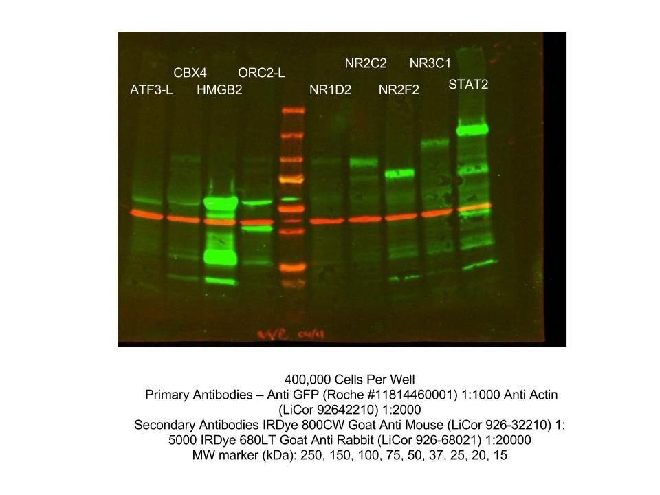
Caption excerpt: Western blot analysis of nuclear lysates prepared from a K562 stable cell line…
- Caption
- Western blot analysis of nuclear lysates prepared from a K562 stable cell line expressin eGFP-ATF3-L using the Roche anti-GFP 11814450001 antibody. The expected MW is 54kDa.
- Submitted by
- Mark Murphy
- Lab
- Kevin White, UChicago
- Grant
- U54HG006996
immunoblot
Caption: immunoblot of GFP tagged ATF3-L, expressed in K562 cells

- Submitted by
- Alec Victorsen
- Lab
- Kevin White, UChicago
- Grant
- U54HG006996
FACs analysis
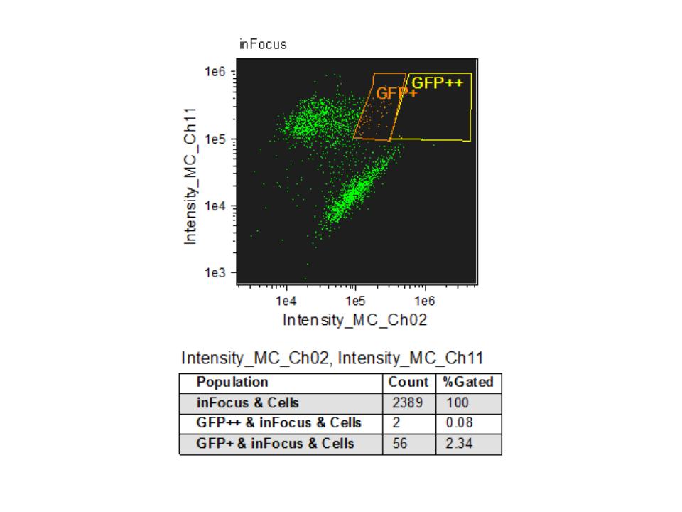
Caption excerpt: eGFP-ATF3 tagged cells were stained with a nuclear dye and sorted by FACS.…
- Caption
- eGFP-ATF3 tagged cells were stained with a nuclear dye and sorted by FACS. Intensities in the GFP channel are on the x-axis, nuclear stain on the y-axis. The boxed regions define a range of intensities in both channels corresponding to GFP+ and GFP++ for the high GFP and highest GFP expression cell populations, respectively. 2389 cells were counted in total, with 2.34% in the GFP+ region and 0.08% in the GFP++ regions respectively. The cells in these regions were further counted for GFP and nuclear signal colocalization and reported as a percentage of GFP+ and GFP++ cells localized to the nucleus: 56.9%. This enriched stable cell line population is then isolated, expanded and stored as frozen aliquots for downstream experiments.
- Submitted by
- Jay Rehm
- Lab
- Kevin White, UChicago
- Grant
- U54HG006996
FACs analysis
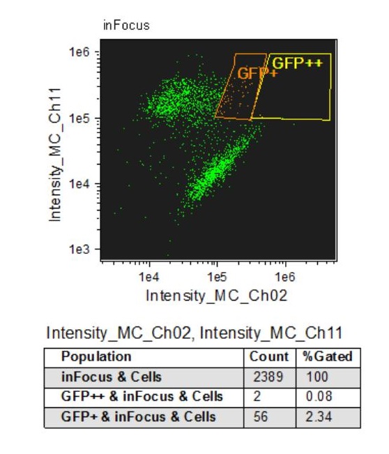
Caption: eGFP-ATF3 tagged cells were stained with a nuclear dye and sorted by FACS.

- Submitted by
- Jay Rehm
- Lab
- Kevin White, UChicago
- Grant
- U54HG006996
FACs analysis
Caption: eGFP-ATF3 tagged cells were stained with a nuclear dye and sorted by FACS.

- Submitted by
- Jay Rehm
- Lab
- Kevin White, UChicago
- Grant
- U54HG006996
immunoblot
Caption excerpt: Western blot analysis of nuclear lysates prepared from a K562 stable cell line…

- Caption
- Western blot analysis of nuclear lysates prepared from a K562 stable cell line expressing eGFP-ATF3 using the Roche anti-GFP 11814450001 antibody. The expected MW is 54kDa.
- Submitted by
- Jay Rehm
- Lab
- Kevin White, UChicago
- Grant
- U54HG006996
FACs analysis
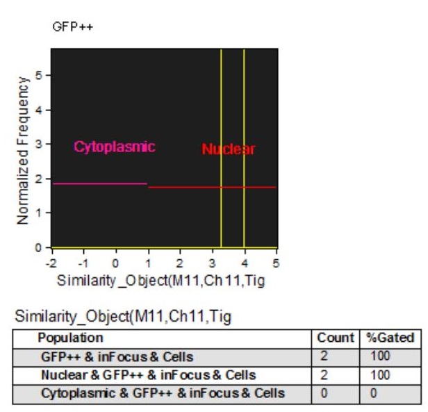
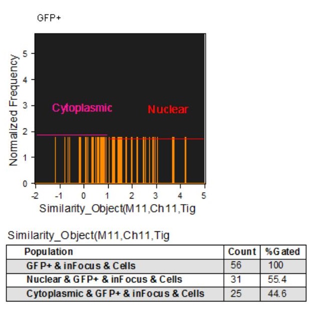
Caption excerpt: eGFP-ATF3-L tagged cells were stained with a nuclear dye, Vybrant DyeCycle…
- Caption
- eGFP-ATF3-L tagged cells were stained with a nuclear dye, Vybrant DyeCycle Ruby Stain from Life technologies, and sorted by FACS. Shown are the cells from the GFP+ fraction quantified for their GFP and nuclear signal colocalization.
- Submitted by
- Mark Murphy
- Lab
- Kevin White, UChicago
- Grant
- U54HG006996
FACs analysis
Caption excerpt: eGFP-ATF3-L tagged cells were stained with a nuclear dye, Vybrant DyeCycle…
- Caption
- eGFP-ATF3-L tagged cells were stained with a nuclear dye, Vybrant DyeCycle Ruby Stain from Life technologies, and sorted by FACS. Shown are the cells from the GFP++ fraction quantified for their GFP and nuclear signal colocalization.
- Submitted by
- Mark Murphy
- Lab
- Kevin White, UChicago
- Grant
- U54HG006996
FACs analysis
Caption excerpt: eGFP-ATF3-L tagged cells were stained with a nuclear dye, Vybrant DyeCycle…
- Caption
- eGFP-ATF3-L tagged cells were stained with a nuclear dye, Vybrant DyeCycle Ruby Stain from Life technologies, and sorted by FACS. Shown are the cells from the GFP+ fraction quantified for their GFP and nuclear signal colocalization.
- Submitted by
- Mark Murphy
- Lab
- Kevin White, UChicago
- Grant
- U54HG006996
general protocol
Description: Protocol for generating GFP tagged BAC transgenic lines in K562.
- Submitted by
- Alec Victorsen
- Lab
- Kevin White, UChicago
- Grant
- U54HG006996