ENCAB995HZA
Antibody against Homo sapiens STAU2
Homo sapiens
HepG2
characterized to standards
Homo sapiens
any cell type or tissue
partially characterized
- Status
- released
- Source (vendor)
- MBLI
- Product ID
- RN013P
- Lot ID
- 001
- Characterized targets
- STAU2 (Homo sapiens)
- Host
- rabbit
- Clonality
- polyclonal
- Purification
- affinity
- Antigen description
- Peptide, 379-396 aa
- Antigen sequence
- TNLQDQLEKTGENKGWSG
- External resources
Characterizations
STAU2 (Homo sapiens)
HepG2
Method: immunoprecipitation
compliant
- Caption
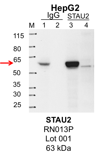
- IP-Western Blot analysis of HepG2 whole cell lysate using STAU2 specific antibody. Lane 1 is 1% of twenty million whole cell lysate input and lane 2 is 25% of IP enrichment using rabbit normal IgG (lanes under 'IgG'). Lane 3 is 1% of twenty million whole cell lysate input and lane 4 is 10% IP enrichment using rabbit polyclonal anti-STAU2 antibody (lanes under 'STAU2').
- Submitted by
- Steven Blue
- Lab
- Gene Yeo, UCSD
- Grant
- U54HG007005
- Download
- HepG2_MBLI_RN013P_001_STAU2.png
STAU2 (Homo sapiens)
HepG2
Method: immunoprecipitation
compliant
- Caption
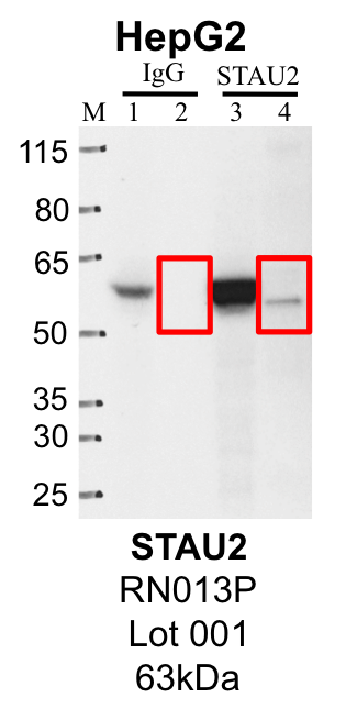
- Representative image of immunoprecipitation performed on whole cell extracts from the HepG2 cell line using the STAU2-specific antibody RN013P. Lane 1: Input from IP using control IgG. Lane 2: Immunoprecipitated material using control IgG. Lane 3: Input from IP using STAU2 antibody. Lane 4: Immunoprecipitated material using STAU2 antibody. Outlined regions were excised from gel and subjected to analysis by mass spectrometry. Target molecular weight: 62.64 kDa.
- Submitted by
- Steven Blue
- Lab
- Gene Yeo, UCSD
- Grant
- U54HG007005
- Download
- HepG2_RN013P_001_STAU2_MassSpec.png
STAU2 (Homo sapiens)
Method: immunoprecipitation
not submitted for review by lab
- Caption
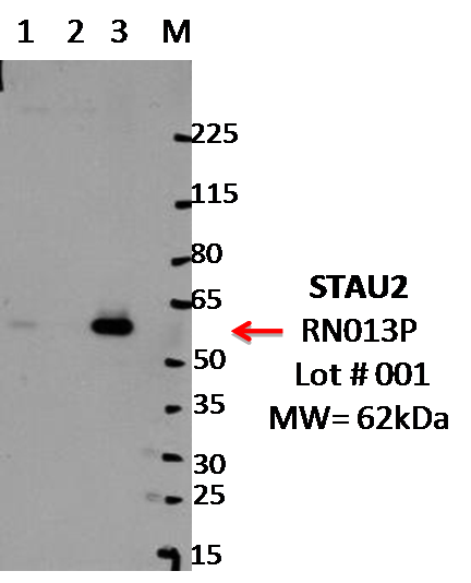
- IP-WB analysis of K562 whole cell lysate using STAU2 specific antibody. Lane 1 is 2.5% of 0.5mg input lysate, lane 2 is 2.5% of supernatant after immunoprecipitation and Lane 3 is 50% of IP enrichment using rabbit polyclonal Anti-STAU2(Human)pAb. This antibody passes preliminary validation and will be further pursued for primary and secondary validation.
- Submitted by
- Balaji Sundararaman
- Lab
- Gene Yeo, UCSD
- Grant
- U54HG007005
- Download
- MBLI_RN013P_001_STAU2.png
STAU2 (Homo sapiens)
Method: immunoprecipitation followed by mass spectrometry
compliant
- Caption
- IP followed by mass spectrometry. Protein was immunoprecipitated from HepG2 whole cell lysates using the antibody RN013P, loaded on a 4-12% NuPAGE Bis-Tris gel, and separated via electrophoresis. Using a reference western blot done in parallel, gel pieces corresponding to the sections indicated were excised and submitted for analysis by the UCSD Biomolecular and Proteomics Mass Spectrometry Facility.
- Submitted by
- Steven Blue
- Lab
- Gene Yeo, UCSD
- Grant
- U54HG007005
- Download
- STAU2 HepG2.pdf
STAU2 (Homo sapiens)
Method: knockdown or knockout
compliant
- Caption
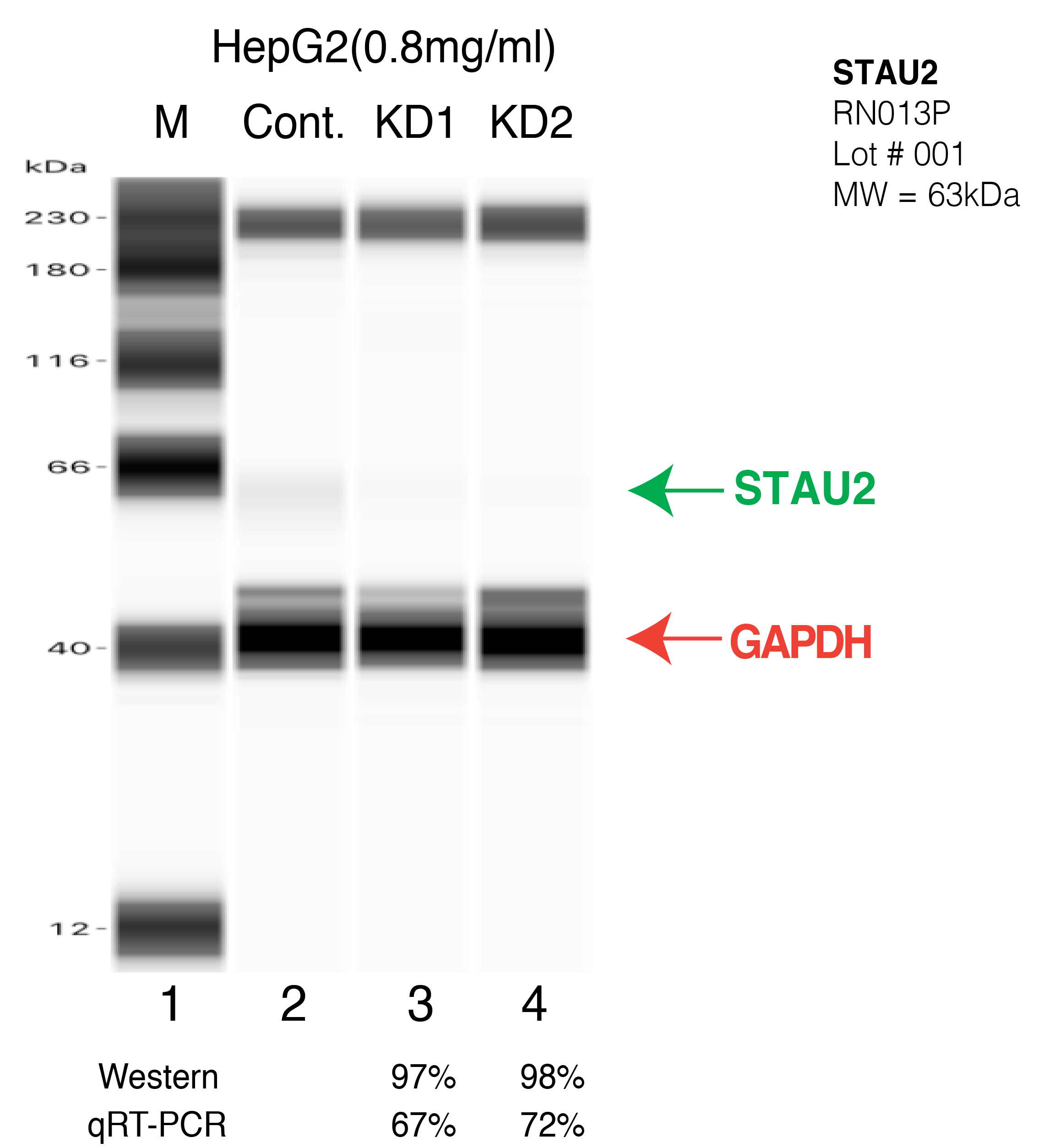
- Western blot following CRISPR against STAU2 in HepG2 whole cell lysate using STAU2 specific antibody. Lane 1 is a ladder, lane 2 is HepG2 non-targeting control knockdown, lane 3 and 4 are two different CRISPR against STAU2.STAU2 protein appears as the green arrow, GAPDH serves as a control and appears in red arrow.
- Submitted by
- Xintao Wei
- Lab
- Brenton Graveley, UConn
- Grant
- U41HG009889
- Download
- STAU2-HEPG2-CRISPR.png
STAU2 (Homo sapiens)
Method: knockdown or knockout
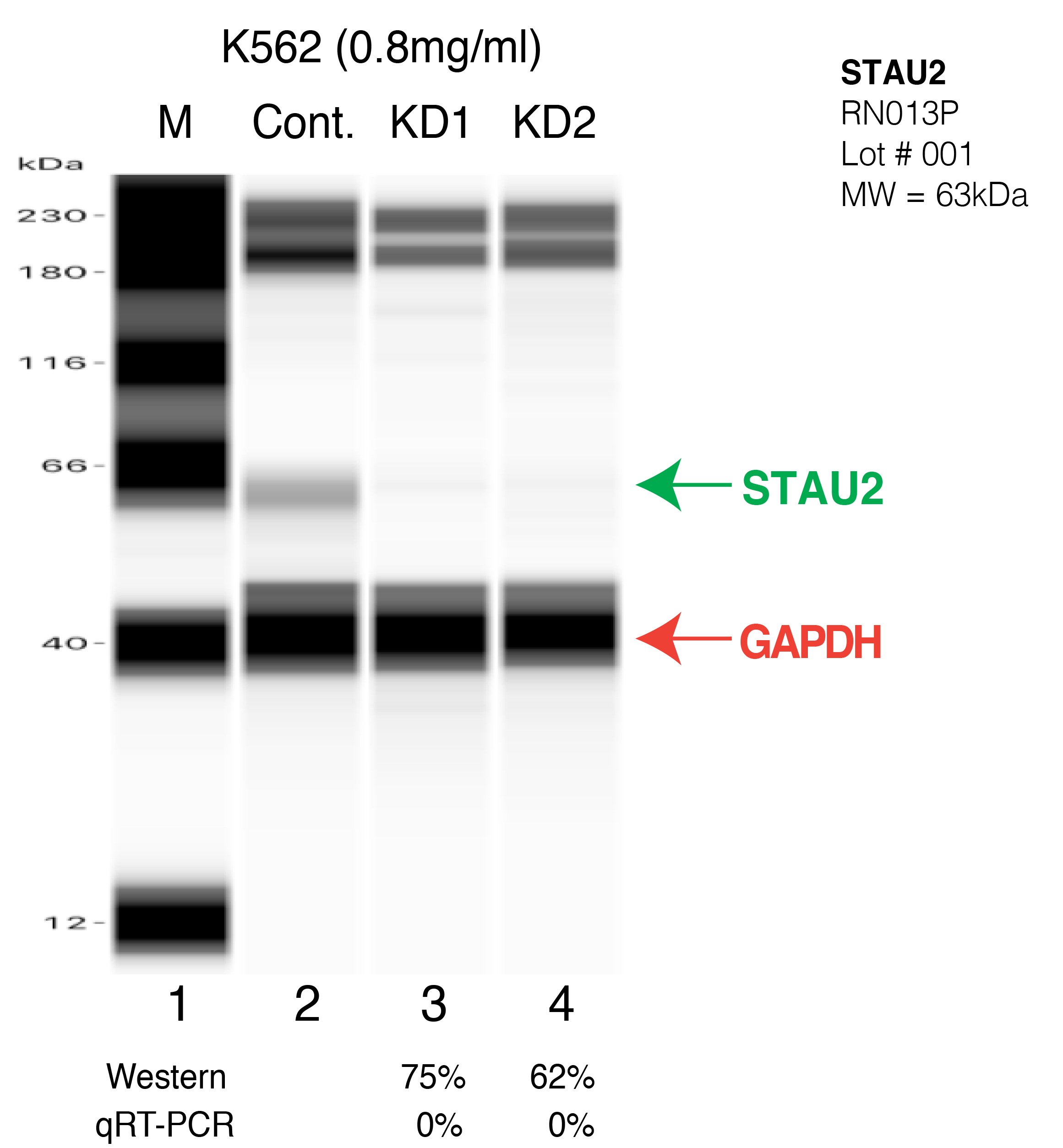
compliant
- Caption
- Western blot following CRISPR against STAU2 in K562 whole cell lysate using STAU2 specific antibody. Lane 1 is a ladder, lane 2 is K562 non-targeting control knockdown, lane 3 and 4 are two different CRISPR against STAU2. STAU2 protein appears as the green arrow, GAPDH serves as a control and appears in red arrow.
- Submitted by
- Xintao Wei
- Lab
- Brenton Graveley, UConn
- Grant
- U41HG009889
- Download
- STAU2-K562-CRISPR.png