ENCAB956LLD
Antibody against Homo sapiens MIER1
Homo sapiens
K562
characterized to standards with exemption
- Status
- released
- Source (vendor)
- Sigma
- Product ID
- HPA019589
- Lot ID
- R08028
- Characterized targets
- MIER1 (Homo sapiens)
- Host
- rabbit
- Clonality
- polyclonal
- Purification
- affinity
- Isotype
- IgG
- Antigen description
- Mesoderm induction early response protein 1 recombinant protein epitope signature tag (PrEST)
- Antigen sequence
- MMEGETNFSSEIEDLAREGDMPIHELLSLYGYGSTVRLPEEDEEEEEEEEEGEDDEDADNDDNSGCSGENKEENIKDSSGQEDETQSSNDDPSQSVASQDAQEI
- Aliases
- michael-snyder:748
- External resources
Characterizations
MIER1 (Homo sapiens)
K562
Method: immunoprecipitation

exempt from standards
- Caption
- Immunoprecipitation of MIER1 from K562 cells using HPA019589. Lane 1: input nuclear lysate. Lane 2: material immunoprecipitated with HPA019589. Lane 3: material immunoprecipitated using control IgG. Band A and BandB were excised from gel and subject to analysis by mass spectrometry. The expected band size is 58 kDa.
- Submitter comment
- Mass spec shows target in band A.
- Reviewer comment
- Multiple bands at higher than expected size, but target confirmed by mass-spec.
- Submitted by
- Kathrina Onate
- Lab
- Michael Snyder, Stanford
- Grant
- U54HG006996
- Download
- 1006_MIER1.jpg
MIER1 (Homo sapiens)
K562
Method: immunoprecipitation
not compliant
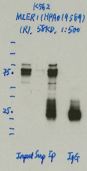
- Caption
- Immunoprecipitation was performed on nuclear extracts from the cell line: K562, using the antibody HPA019589. The blot shows western blot analysis of input, flowthrough, immunoprecipitate and mock immunoprecipitate using IgG.
- Reviewer comment
- too many bands, not >50% of total signal
- Submitted by
- Denis Salins
- Lab
- Michael Snyder, Stanford
- Grant
- U54HG006996
MIER1 (Homo sapiens)
Method: immunoprecipitation followed by mass spectrometry
compliant
- Caption
- IP followed by mass spectrometry: Briefly, protein was immunoprecipitated from K562 nuclear cell lysates using HPA019589, and the IP fraction was loaded on a 10% polyacrylamide gel (NuPAGEBis-Tris Gel) and separated with an Invitrogen NuPAGE electrophoresis system. The gel was stained by ColloidialCoomassie G-250 stain, gel fragments corresponding to the bands indicated were excised. Then proteins were trypsinized using the in-gel digestion method. Digested proteins were analyzed on an Orbitrap Elite mass spectrometer (Thermo Scientific) by the nanoLC-ESI-MS/MS technique. Peptides were identified by the SEQUEST algorithm and filtered with a high confidence threshold (Peptide false discovery rate < 1%, 2 unique peptides per protein minimum, mass error < 10 ppm).
- Reviewer comment
- BAHD1 has same unique peptide count and is also DNA binding. Do they interact?
- Submitted by
- Kathrina Onate
- Lab
- Michael Snyder, Stanford
- Grant
- U54HG006996