ENCAB937ZMF
Antibody against Homo sapiens REST
Homo sapiens
K562, HepG2, GM12878, endothelial cell of umbilical vein, H1, A549
characterized to standards with exemption
- Status
- released
- Source (vendor)
- Millipore
- Product ID
- 17-641
- Lot ID
- 2384024
- Characterized targets
- REST (Homo sapiens)
- Host
- rabbit
- Clonality
- polyclonal
- Antigen description
- RE1-silencing transcription factor
- Aliases
- bradley-bernstein:PchAb 1223
- External resources
Characterizations
REST (Homo sapiens)
H1A549
Method: immunoblot

exempt from standards
- Submitter comment
- --
- Reviewer comment
- --
- Submitted by
- Nina Farrell
- Lab
- Bradley Bernstein, Broad
- Grant
- U54HG006991
- Download
- exempted.png
REST (Homo sapiens)
Method: ChIP-seq comparison
exempt from standards
- Caption
- REST is a sequence specific binding factor. As such, the validation standard for REST antibodies is distinct from the chromatin regulator validation standard under which the remainder of our antibody validation dossiers are being constructed. The present document does not intend to substitute for the full computational analysis for the enrichment of the recognition sequence for REST in peaks discovered using ChIP-seq. The present document will merely illustrate the tracks we have obtained using two distinct lots of a particular antibody, and will present a single illustration of the occurrence of a perfect occurrence of the REST recognition sequence in tight proximity to a ChIP-seq peak, using a particular locus [CHRNB2 (cholinergic receptor nicotinic beta 2 subunit)] identified in the classic literature on the genome-wide occurrence of the recognition motif of the neuron-restrictive silencing factor (REST). Many thousands of other such illustrations could also be presented.
- Submitter comment
- This was cleared by the antibody committee
- Reviewer comment
- This is a well characterized antibody already used in many ENCODE experiments.
- Submitted by
- Nina Farrell
- Lab
- Bradley Bernstein, Broad
- Grant
- U54HG006991
- Download
- REST PchAb 448 WIP SAV.docx.pdf
REST (Homo sapiens)
K562HepG2GM12878endothelial cell of umbilical vein
Method: immunoblot
compliant
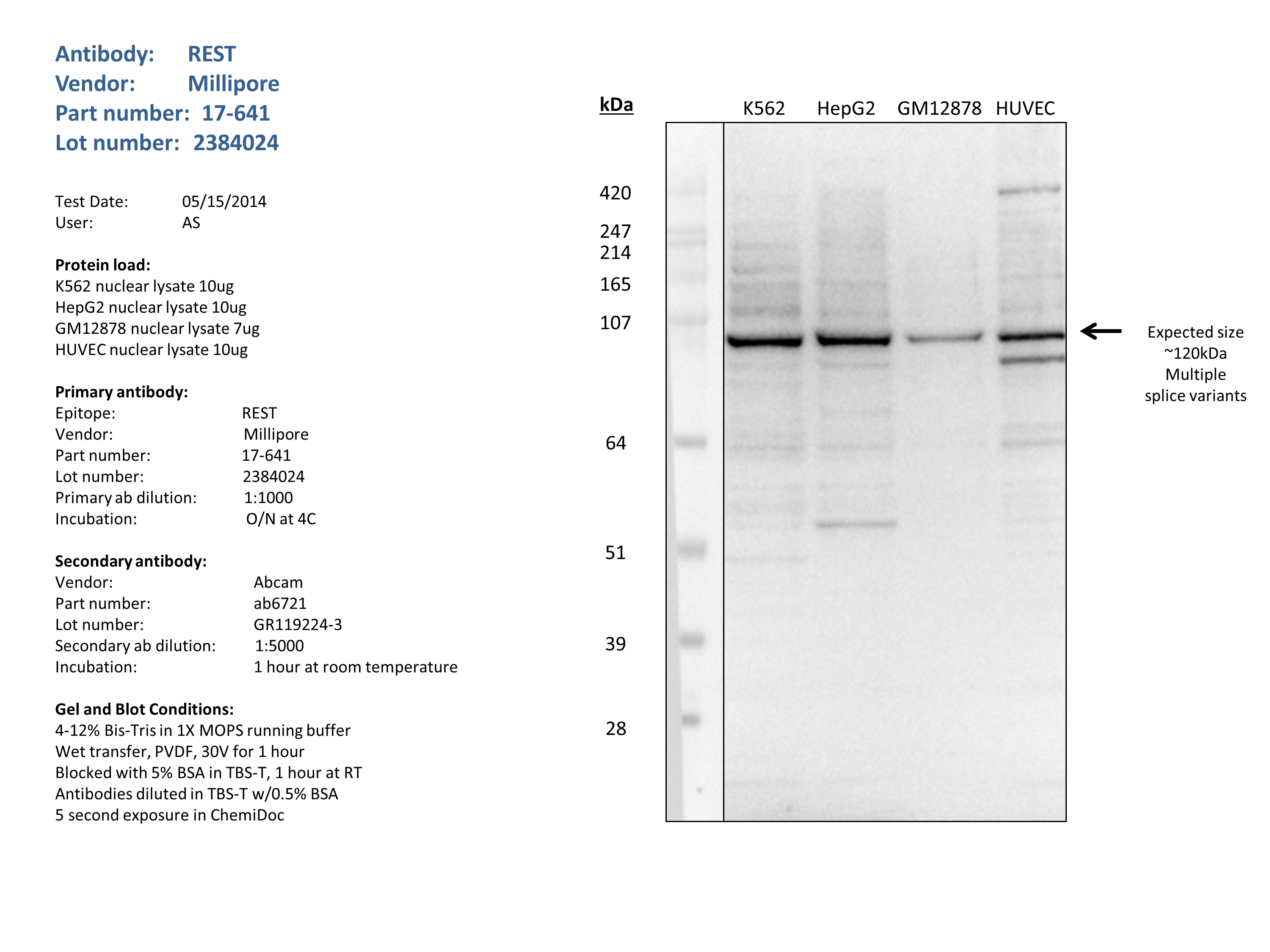
- Caption
- Nuclear lysates from K562 (10ug), HepG2 (10ug), GM12878 (7ug), HUVEC (10ug), were loaded into a 4-12% Bis-Tris gel in 1X MOPS running buffer. After separation, the samples were transferred to a PVDF membrane using wet transfer. Membrane was blocked for an hour in room temperature, with 5% BSA in TBS-T and blotted with primary antibody in the appropriate concentration over night at 4c. Membrane was washed and blotted with secondary HRP-conjugated antibody. Detection was made with Optiblot ECL Detect Kit (ab133406) for 2 min. The strongest band was detected around the expected size (~120kDa).
- Submitted by
- Noam Shoresh
- Lab
- Bradley Bernstein, Broad
- Grant
- U54HG006991
- Download
- REST_Millipore_17-641_2384024.png