ENCAB741YHG
Antibody against Homo sapiens SUZ12
Homo sapiens
HepG2, endothelial cell of umbilical vein
characterized to standards
Homo sapiens
GM12878
characterized to standards with exemption
- Status
- released
- Source (vendor)
- Cell Signaling
- Product ID
- 3737BF
- Lot ID
- 4
- Characterized targets
- SUZ12 (Homo sapiens)
- Host
- rabbit
- Clonality
- monoclonal
- Antigen description
- Sites of PcG activity
- Aliases
- bradley-bernstein:PchAb 1147
- External resources
Characterizations
SUZ12 (Homo sapiens)
GM12878
Method: immunoblot

exempt from standards
- Caption
- This antibody was used before a characterization for GM12878 was performed. It has been reviewed and exempted by the antibody characterization board.
- Submitter comment
- The antibody has a conforming western only in HepG2 and HUVEC. A third dataset (https://www.encodeproject.org/experiments/ENCSR091BOQ/) used this antibody in GM12878. Release of that single dataset is blocked due to the lack of primary validation in that cell type. We would like an exception to allow the release of this one dataset. We do not anticipate further use of this antibody and have no remaining inventory.
- Reviewer comment
- PJF: I agree that this dataset should be released without requiring a western blot in GM12978 (using whatever term is appropriate such as “characterized to standard with exemption”).
- Submitted by
- Nina Farrell
- Lab
- Bradley Bernstein, Broad
- Grant
- U54HG006991
- Download
- exempted.png
SUZ12 (Homo sapiens)
Method: ChIP-seq comparison
compliant
- Caption
- This validation relies on the use of antibodies to different members of a known complex, and the demonstration that highly similar patterns of enrichment are obtained with each antibody. The second track shown used an antibody to EZH2 (PchAb 58-V), another member of the PRC2 complex. Overall correlation score: SUZ12 vs EZH2: 0.9439.
- Submitted by
- Kathrina Onate
- Lab
- Bradley Bernstein, Broad
- Grant
- U54HG006991
- Download
- SUZ12 CST 3737BF lot 4 SAV (1).pdf
SUZ12 (Homo sapiens)
HepG2endothelial cell of umbilical vein
Method: immunoblot
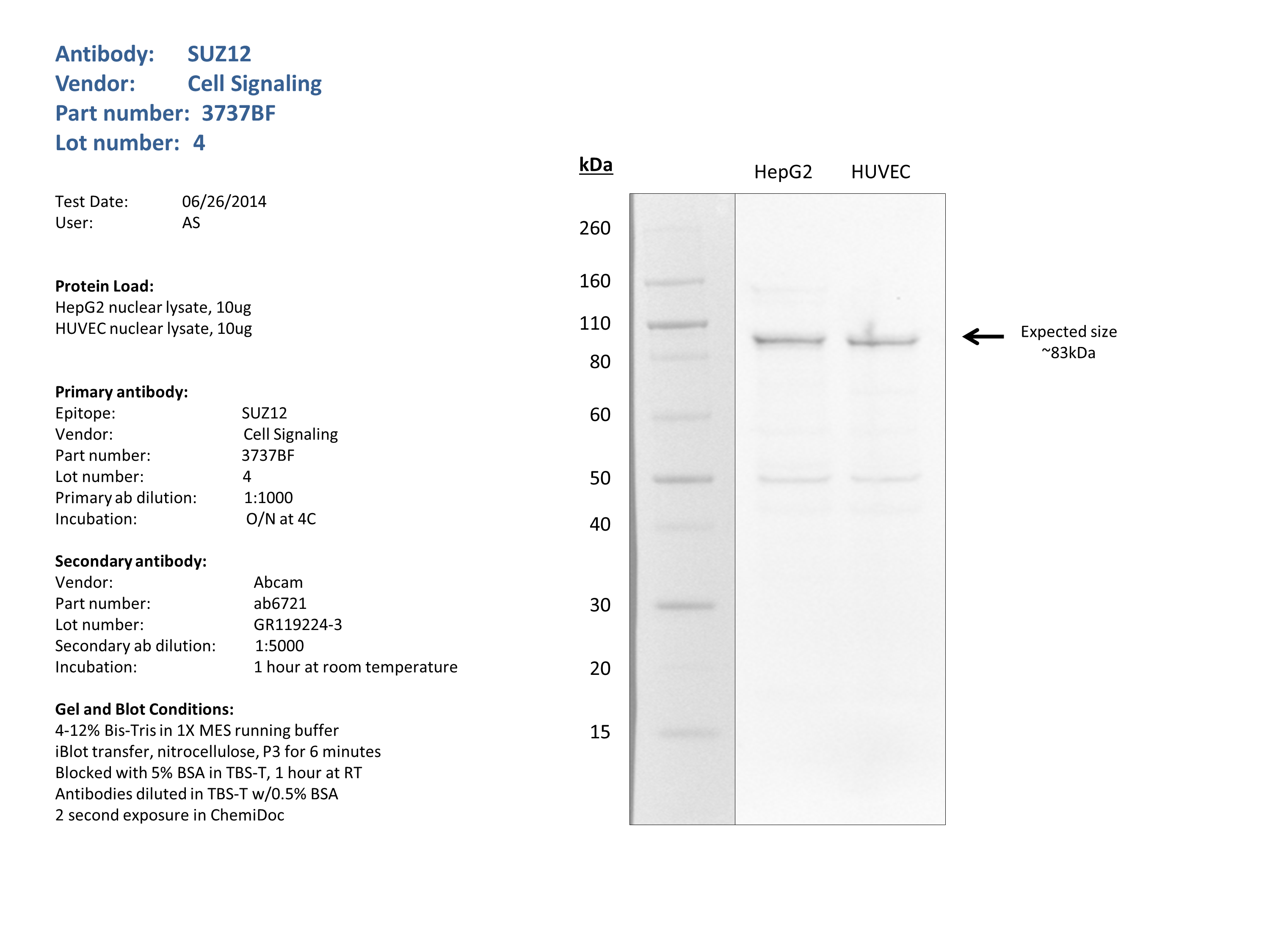
compliant
- Caption
- Nuclear lysates from HepG2 (10ug), HUVEC (10ug), were loaded into a 4-12% Bis-Tris gel in 1X MES running buffer. After separation, the samples were transferred to a nitrocellulose membrane using the iblot system. Membrane was blocked for an hour in room temperature, with 5% BSA in TBS-T and blotted with primary antibody in the appropriate concentration over night at 4c. Membrane was washed and blotted with secondary HRP-conjugated antibody. Detection was made with Optiblot ECL Detect Kit (ab133406) for 2 min.A band of the expected size was detected (~83kDa).
- Submitted by
- Noam Shoresh
- Lab
- Bradley Bernstein, Broad
- Grant
- U54HG006991