ENCAB324ARN
Antibody against Homo sapiens NR2C1
Homo sapiens
K562, MCF-7, GM12878, HepG2
characterized to standards with exemption
- Status
- released
- Source (vendor)
- Bethyl Labs
- Product ID
- A303-047A
- Lot ID
- 1
- Characterized targets
- NR2C1 (Homo sapiens)
- Host
- rabbit
- Clonality
- polyclonal
- Purification
- affinity
- Aliases
- michael-snyder:AS-1275
- External resources
Characterizations
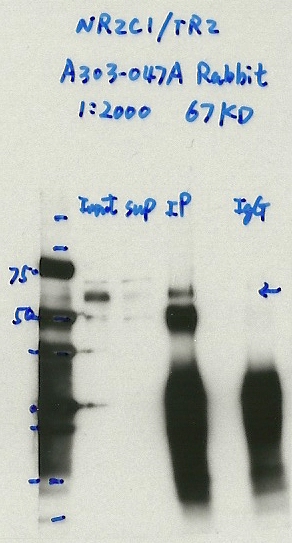
NR2C1 (Homo sapiens)
K562
Method: immunoprecipitation
exempt from standards
- Caption
- Immunoprecipitation was performed on nuclear extracts from the cell line: K562, using the antibody A303-047A. The blot shows western blot analysis of input, flowthrough, immunoprecipitate and mock immunoprecipitate using IgG. Expected Molecular Weight 67 KD
- Submitter comment
- -
- Reviewer comment
- Since both bands disappear with IgG, I think they both are the protein
- Submitted by
- Denis Salins
- Lab
- Michael Snyder, Stanford
- Grant
- U54HG006996
- Download
- Expt1055_6-NR2C1_TR2_A303-047A.jpg
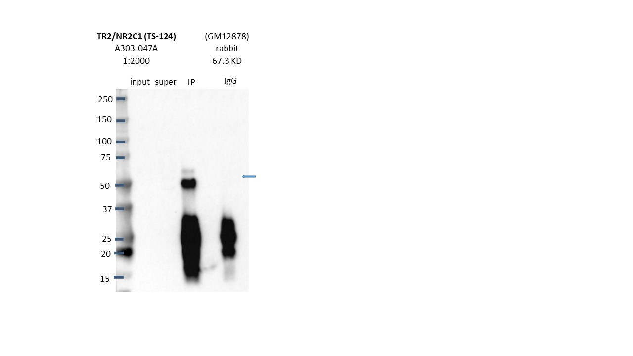
NR2C1 (Homo sapiens)
GM12878
Method: immunoprecipitation
exempt from standards
- Caption
- Immunoprecipitation was performed on nuclear extracts from the cell line: GM12878 using the antibody A303-047A. The image shows western blot analysis of input, flowthrough, immunoprecipitate, and mock immunoprecipitate using IgG. Target molecular weight: 67.315.
- Submitter comment
- 1) same pattern as K562 IP. 2) Mass Spec result indicates that the both bands (~50-75kd) are NR2C1
- Reviewer comment
- Marked band at expected size not 50% of total signal in lane is consistent with banding pattern in K562 which was analyzed by mass spec and confirmed to detect NR2C1.
- Submitted by
- Nathaniel Watson
- Lab
- Michael Snyder, Stanford
- Grant
- U54HG006996
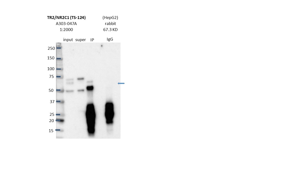
NR2C1 (Homo sapiens)
HepG2
Method: immunoprecipitation
exempt from standards
- Caption
- Immunoprecipitation was performed on nuclear extracts from the cell line: HepG2 using the antibody A303-047A. The image shows western blot analysis of input, flowthrough, immunoprecipitate, and mock immunoprecipitate using IgG. Target molecular weight: 67.315.
- Submitter comment
- 1) same pattern as K562 IP. 2) Mass Spec result indicates that the both bands (~50-75kd) are NR2C1
- Reviewer comment
- Marked band at expected size not 50% of total signal in lane is consistent with banding pattern in K562 which was analyzed by mass spec and confirmed to detect NR2C1.
- Submitted by
- Nathaniel Watson
- Lab
- Michael Snyder, Stanford
- Grant
- U54HG006996
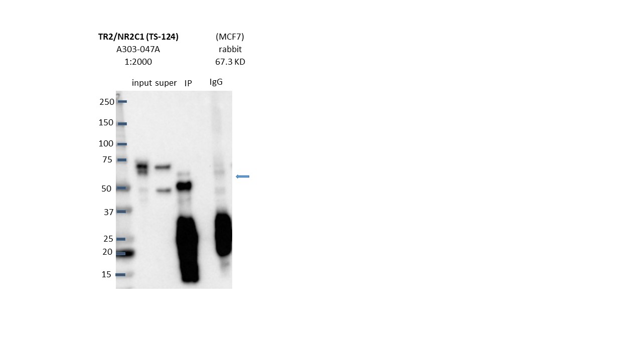
NR2C1 (Homo sapiens)
MCF-7
Method: immunoprecipitation
exempt from standards
- Caption
- Immunoprecipitation was performed on nuclear extracts from the cell line: MCF-7 using the antibody A303-047A. The image shows western blot analysis of input, flowthrough, immunoprecipitate, and mock immunoprecipitate using IgG. Target molecular weight: 67.315.
- Submitter comment
- 1) same pattern as K562 IP. 2) Mass Spec result indicates that the both bands (~50-75kd) are NR2C1
- Reviewer comment
- Marked band at expected size not 50% of total signal in lane is consistent with banding pattern in K562 which was analyzed by mass spec and confirmed to detect NR2C1.
- Submitted by
- Nathaniel Watson
- Lab
- Michael Snyder, Stanford
- Grant
- U54HG006996
NR2C1 (Homo sapiens)
K562
Method: immunoprecipitation
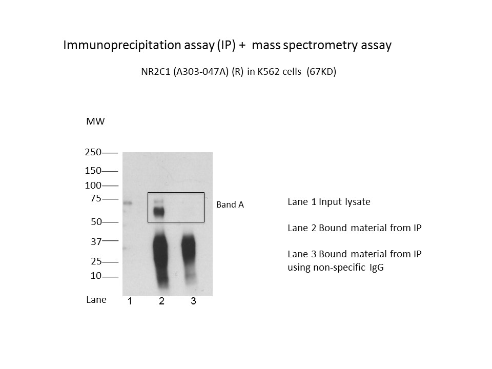
exempt from standards
- Caption
- Immunoprecipitation was performed on nuclear extracts from the cell line K562 using the antibody A303-047A. Lane 1: input nuclear lysate. Lane 2: material immunoprecipitated with antibody. Lane 3: material immunoprecipitated using control IgG. Marked bands were excised from gel and subjected to analysis by mass spectrometry. Target molecular weight: 67.315.
- Submitter comment
- -
- Reviewer comment
- Cut section analyzed by mass-spec encompasses two immunoreactive bands analyzed by mass-spec, but banding pattern is consistent with vendor's IPs. Mass-spec detected NR2C1
- Submitted by
- Nathaniel Watson
- Lab
- Michael Snyder, Stanford
- Grant
- U54HG006996
- Download
- NR2C1(A303-047A).jpg
NR2C1 (Homo sapiens)
Method: immunoprecipitation followed by mass spectrometry
compliant
- Caption
- IP followed by mass spectrometry. Briefly, protein was immunoprecipitated from K562 nuclear cell lysates using the antibody A303-047A, and the IP fraction was loaded on a 10% polyacrylamide gel (NuPAGEBis-Tris Gel) and separated with an Invitrogen NuPAGE electrophoresis system. The gel was stained by ColloidialCoomassie G-250 stain, gel fragments corresponding to the bands indicated were excised. Then proteins were trypsinized using the in-gel digestion method. Digested proteins were analyzed on an Orbitrap Elite mass spectrometer (Thermo Scientific) by the nanoLC-ESI-MS/MS technique. Peptides were identified by the SEQUEST algorithm and filtered with a high confidence threshold (Peptide false discovery rate < 1%, 2 unique peptides per protein minimum, mass error < 10 ppm).
- Submitter comment
- CCAR1, FUS and DLAT are not sequence-specific DNA binding TFs.
- Submitted by
- Nathaniel Watson
- Lab
- Michael Snyder, Stanford
- Grant
- U54HG006996
- Download
- NR2C1_A303-047A_final.pdf