ENCAB000BKT
Antibody against Homo sapiens H3K36me3
Homo sapiens
any cell type or tissue
characterized to standards
- Status
- released
- Source (vendor)
- Abcam
- Product ID
- ab9050
- Lot ID
- 446805
- Characterized targets
- H3K36me3 (Homo sapiens)
- Host
- rabbit
- Clonality
- polyclonal
- Aliases
- bradley-bernstein:PchAb 457
- External resources
Characterizations
H3K36me3 (Homo sapiens)
Method: ChIP-seq comparison
compliant
- Caption
- We used Pearson Correlation to compare the two tracks. Metrics were derived as follows: (1) Genomic windows for validation test: We collated a set of 15,157 3KB genomic windows whose ChIP-seq signals vary between histone marks and cell types, based on a manually curated set of ~1000 ChIP-seq experiments (data from the Encode2 and NIH Roadmap Epigenomics projects [URL here]). The genomic coordinates of these 15,157 3KB windows are available here. For the current validation exercise, we excluded 500 windows with false-positive constitutive signals across marks and input controls. (2) Antibody validation test: For each of the two ChIP-seq experiments, numerical values corresponding to normalized read density for each window were calculated. The Pearson correlation between both the two tracks was computed using the retained 14,657 genomic intervals and is reported above.
- Submitter comment
- I would be happy to give this a “complaint” based on the western blot as primary and comparison to the other dataset as secondary. Peggy Farnham November 11th, 2016
- Reviewer comment
- This antibody is being validated by comparison to ENCAB000ADU (Broad alias PchAb 78-V).
- Submitted by
- Noam Shoresh
- Lab
- Bradley Bernstein, Broad
- Grant
- U54HG006991
H3K36me3 (Homo sapiens)
K562
Method: immunoblot
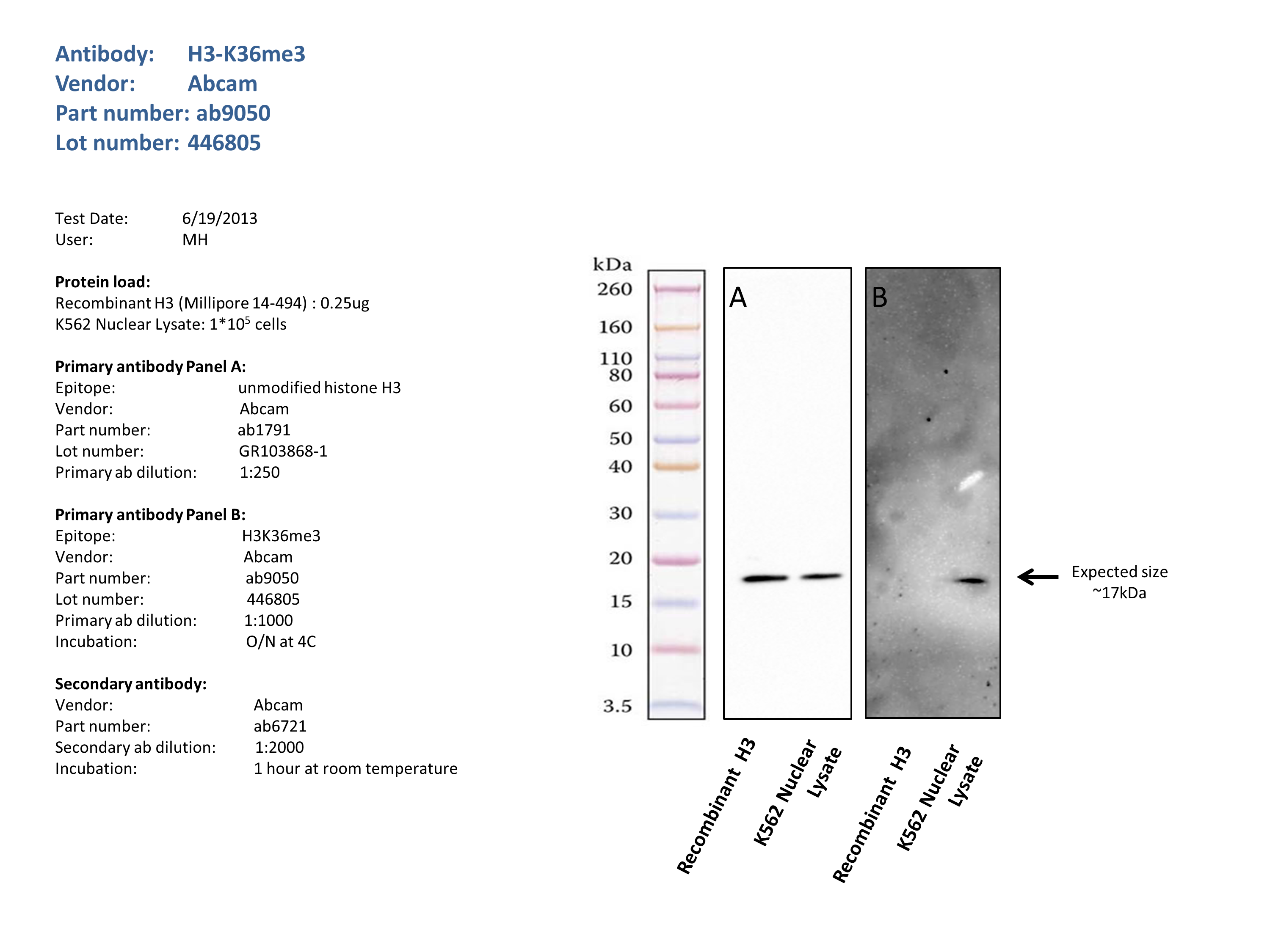
compliant
- Caption
- 0.25ug unmodified Recombinant Histone H3 (Millipore 14-494) and 30ug of K562 Whole Cell Lysate (Abcam, ab7911) were resolved by electrophoresis on a 4-12% acrylamide gel. After separation, the samples were transferred to a nitrocellulose membrane with an Invitrogen iBlot system. Membrane was blocked for an hour in room temperature, with 5% nonfat dry milk and blotted with primary antibody in the appropriate concentration over night at 4c. Membrane was washed and blotted with secondary HRP-conjugated antibody. Detection was made with Optiblot ECL Detect Kit (ab133406) for 2 min. Panel A: anti Histone H3 (Abcam ab1791), band of expected size (~17kDa) visualized in both unmodified recombinant histone H3 and WCL, at similar intensities.Panel B: Band of expected size (~17kDa) visualized in WCL, and cannot be detected in unmodified recombinant Histone H3. Antibody seems to be specific since no reactivity with nonhistone proteins or unmodified histones was detected
- Submitted by
- Noam Shoresh
- Lab
- Bradley Bernstein, Broad
- Grant
- U54HG006991