ENCAB000BDZ
Alternate accession: ENCAB738UYY
Antibody against Homo sapiens KAT2B
Homo sapiens
GM12878, K562, HeLa-S3, HepG2, IMR-90, MCF-7
characterized to standards
Homo sapiens
heart
not characterized to standards
- Status
- released
- Source (vendor)
- Cell Signaling
- Product ID
- 3378S
- Lot ID
- 1
- Characterized targets
- KAT2B (Homo sapiens)
- Host
- rabbit
- Clonality
- monoclonal
- Purification
- affinity
- Isotype
- IgG
- Antigen description
- Synthetic peptide corresponding to the amino terminus of human PCAF protein.
- Aliases
- bradley-bernstein:PchAb 1213, michael-snyder:AS-362
- External resources
Characterizations
KAT2B (Homo sapiens)
MCF-7
Method: immunoprecipitation
compliant
- Caption
- Immunoprecipitation was performed on nuclear extracts from the cell line: MCF-7, using the antibody 3378S. The blot shows western blot analysis of input, flowthrough, immunoprecipitate and mock immunoprecipitate using IgG. Molecular weight: 93013 Da
- Submitted by
- Denis Salins
- Lab
- Michael Snyder, Stanford
- Grant
- U54HG006996
- Download
- expt1032_5.jpg
KAT2B (Homo sapiens)
K562
Method: immunoprecipitation
compliant
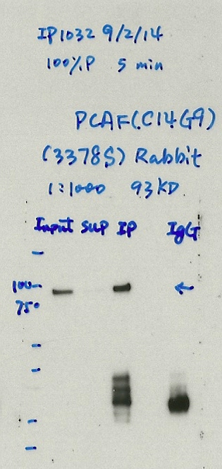
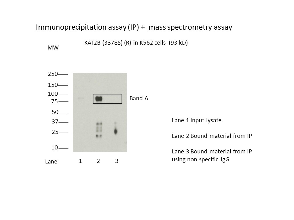
- Caption
- Immunoprecipitation of KAT2B (also known as PCAF) from K562 cells using 3378S. Lane 1: input nuclear lysate. Lane 2: material immunoprecipitated with 3378S. Lane 3: material immunoprecipitated using control IgG. Band A was excised from gel and subject to analysis by mass spectrometry. The expected band size is 93 kDa.
- Submitted by
- Kathrina Onate
- Lab
- Michael Snyder, Stanford
- Grant
- U54HG006996
- Download
- KAT2B(3378S).JPG
KAT2B (Homo sapiens)
Method: immunoprecipitation followed by mass spectrometry
compliant
- Caption
- IP followed by mass spectrometry: Briefly, protein was immunoprecipitated from K562 nuclear cell lysates using 3378S, and the IP fraction was loaded on a 10% polyacrylamide gel (NuPAGEBis-Tris Gel) and separated with an Invitrogen NuPAGE electrophoresis system. The gel was stained by ColloidialCoomassie G-250 stain, gel fragments corresponding to the bands indicated were excised. Then proteins were trypsinized using the in-gel digestion method. Digested proteins were analyzed on an Orbitrap Elite mass spectrometer (Thermo Scientific) by the nanoLC-ESI-MS/MS technique. Peptides were identified by the SEQUEST algorithm and filtered with a high confidence threshold (Peptide false discovery rate < 1%, 2 unique peptides per protein minimum, mass error < 10 ppm).
- Submitter comment
- TAF5L and SUPT20H were detected with more peptides than KAT2B. KAT2B has unique interaction with TAF5L and TAF6L. http://thebiogrid.org/114375/summary/homo-sapiens/kat2b.html SUPT20H has interaction with TAF6L. http://thebiogrid.org/120729/summary/homo-sapiens/supt20h.html
- Submitted by
- Kathrina Onate
- Lab
- Michael Snyder, Stanford
- Grant
- U54HG006996
- Download
- KAT2B_3378S final KAT2B_3378S.pdf
KAT2B (Homo sapiens)
GM12878K562HeLa-S3HepG2IMR-90heart
Method: immunoblot
compliant
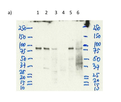
- Caption
- a) Western blot analysis of nuclear lysates prepared from multiple cells lines loaded in the order: GM12878, K562, Hela S3, HepG2, IMR90, A549, Heartusing the antibody 3378S.
- Submitted by
- Trupti Kawli
- Lab
- Michael Snyder, Stanford
- Grant
- U54HG006996
- Download
- KAT2B_3378S_WB_a.jpg
KAT2B (Homo sapiens)
K562
Method: immunoprecipitation
compliant
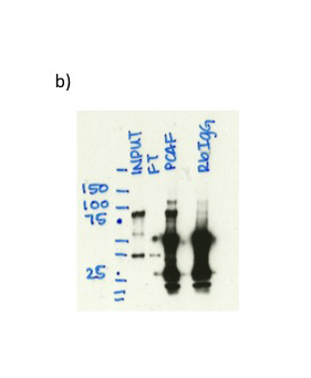
- Caption
- b) Immunoprecipitation was performed on nuclear extracts from the cell line: K562 using the antibody 3378S. The blot shows western blot analysis of input, flowthrough, immunoprecipitate and mock immunoprecipitate using IgG.
- Reviewer comment
- Multiple bands, higher one seems non-specific but band at expected size >50% of total signal in the lane.
- Submitted by
- Trupti Kawli
- Lab
- Michael Snyder, Stanford
- Grant
- U54HG006996
- Download
- KAT2B_3378S_WB_b.jpg
KAT2B (Homo sapiens)
HepG2
Method: immunoprecipitation
compliant
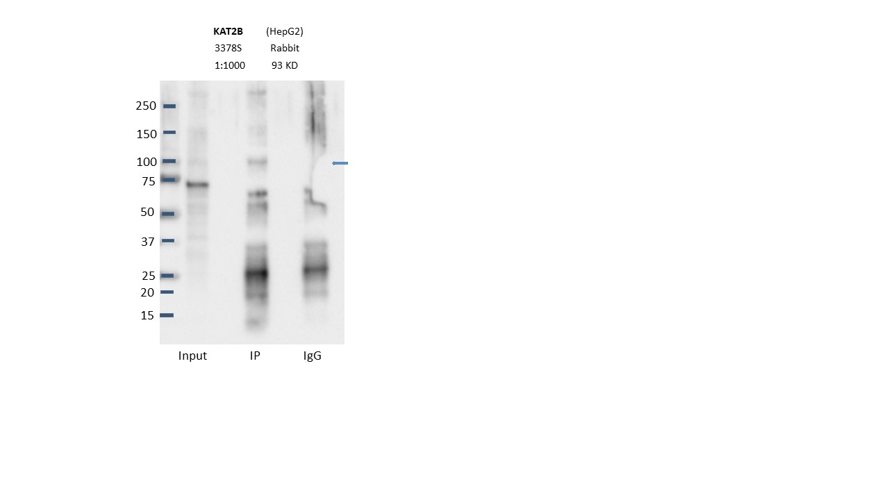
- Caption
- Immunoprecipitation was performed on nuclear extracts from the cell line: HepG2, using the antibody 3378S. The blot shows western blot analysis of input, flowthrough, immunoprecipitate and mock immunoprecipitate using IgG.Molecular Weight: 93.0
- Submitted by
- Nathaniel Watson
- Lab
- Michael Snyder, Stanford
- Grant
- U54HG006996
- Download
- PCAF.JPG
KAT2B (Homo sapiens)
K562
Method: immunoprecipitation
not compliant
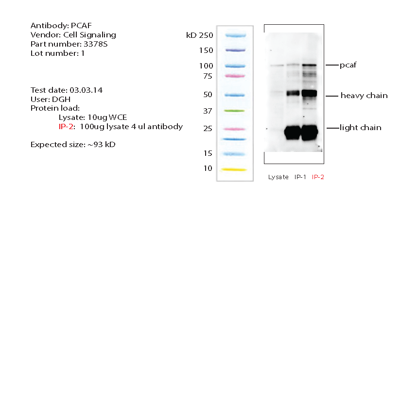
- Caption
- K562 whole cell lysate was immunoprecipitated using primary antibody, and the IP fraction along side a whole cell lysate were loaded on a CriterionXT gel and separated. After separation, the samples were transferred using a wet transfer. Blotting with primary (same as that used for IP) and secondary HRP-conjugated antibodies was performed. Band of expected size visualized representing strongest signal in the lane.
- Reviewer comment
- Missing IgG control.
- Submitted by
- Noam Shoresh
- Lab
- Bradley Bernstein, Broad
- Grant
- U54HG006991
- Download
- pcaf_bethyl_3378S_1_IP.png