ENCAB000AKY
Antibody against Homo sapiens SP1
Homo sapiens
K562, GM12878, liver
characterized to standards with exemption
Homo sapiens
HepG2, MCF-7
not characterized to standards
- Status
- released
- Source (vendor)
- Santa Cruz Biotech
- Product ID
- sc-17824
- Lot ID
- K1907
- Characterized targets
- SP1 (Homo sapiens)
- Host
- mouse
- Clonality
- monoclonal
- Antigen description
- Raised against amino acids 121-345 mapping near the N-terminus of Sp1 of human origin.
- External resources
Characterizations
SP1 (Homo sapiens)
K562GM12878HepG2
Method: immunoprecipitation
exempt from standards
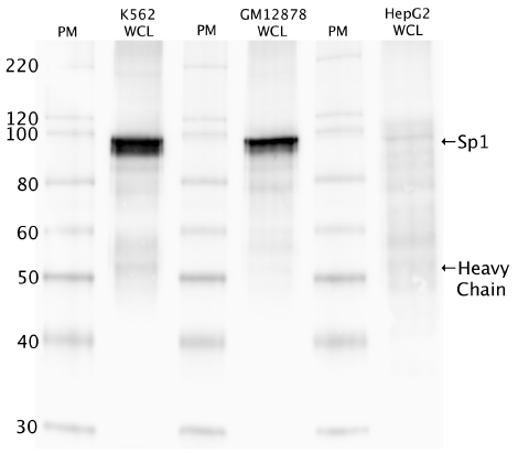
- Caption
- Western blot protocol: Whole cell lysate was immunoprecipitated using primary antibody, and the IP fraction was loaded on a 12% acrylamide gel and separated with a Bio-Rad PROTEAN II xi system. After separation, the samples were transferred to a nitrocellulose membrane with an Invitrogen iBlot system. Blotting with primary (same as that used for IP) and secondary HRP-conjugated antibodies was performed on an Invitrogen BenchPro 4100 system. Visualization was achieved using SuperSignal West Femto solution (Thermo Scientific). Results: Band of expected size visualized, representing strongest signal in the lane. Figure legend: IP-western with sc-17824 in whole cell lysate (WCL) of K562, GM12878 and HepG2; PM=protein marker. SP1 bands are indicated.
- Submitter comment
- The lab is asking for an exemption for this characterization because it was done before the agreements to have an IgG control
- Reviewer comment
- As per Antibody review panal decsion of Feb 29, 2016, this will be exempted from standards
- Submitted by
- Richard Myers
- Lab
- Richard Myers, HAIB
- Grant
- U54HG004576
- Download
- IP_human_SP1_validation_Myers.png
SP1 (Homo sapiens)
liver
Method: immunoblot

exempt from standards
- Caption
- The ENCODE Binding Working Group finds for some valuable tissues that recreating a primary on well characterized antibodies is not cost effective. Therefore, they allow exemption from standards for these tissues.
- Submitter comment
- The lab is asking for an exemption for liver cells due to the lack of resource to make a primary characterization for them
- Reviewer comment
- Exempted by the Feb 29, 2016 antibody review panel
- Submitted by
- Richard Myers
- Lab
- Richard Myers, HAIB
- Grant
- U54HG006998
- Download
- No_tissue.png
SP1 (Homo sapiens)
Method: immunoprecipitation followed by mass spectrometry
exempt from standards
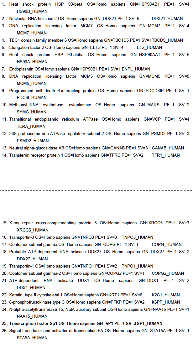
- Caption
- IP followed by mass spectrometry: Briefly, K562 whole cell lysates were immunoprecipitated using primary antibody, and the IP fraction was loaded on a 12% acrylamide gel and separated with a Bio-Rad PROTEAN II xi system. Gel was stained with Coomassie Blue in order to visualize marker bands. Gel fragments corresponding to the bands indicated above in the western blot image were excised and sent to the University of Alabama at Birmingham Cancer Center Mass Spectrometry/Proteomics Shared Facility. There the samples were run on an LTQ XL Linear Ion Trap Mass Spectrometer by LC-ESI-MS/MS. Peptides were identified using SEQUEST tandem mass spectra analysis, with probability based matching at p < 0.05. As per ENCODE data standards, all SEQUEST results are attached (ENCODE_HAIB_SP1_sc17824_09122011_MassSpec.pdf), including common contaminants. Target protein is listed as hit 25 in ~100 kDa band. ENCODE data standards recognizes various methodologies for secondary validation of antibodies. Among these methodologies is immunoprecipitation followed by mass spectrometry analysis. Briefly, K562 whole cell lysates were immunoprecipitated using primary antibody, and the IP fraction was loaded on a 12% acrylamide gel and separated with a Bio-Rad PROTEAN II xi system. Gel was stained with Coomasie Blue in order to visualize marker bands. A gel fragment corresponding to the band indicated above in the western blot image was excised and sent to the University of Alabama at Birmingham Cancer Center Mass Spectrometry/Proteomics Shared Facility. There the sample was run on an LTQ XL Linear Ion Trap Mass Spectrometer with alternating collision-induced dissociation and electron-transfer dissociation. Peptides were identified using MASCOT (Matrix Science), with probability based matching at p < 0.05. Subsequent analysis was performed in Scaffold (Proteome Software, Inc.) at 0.0% protein FDR and 0.0% peptide FDR. As per ENCODE data standards, all Scaffold results are listed below, including common contaminants. Target protein is highlighted in bold font.
- Submitter comment
- This work was done in ENCODE2 before the standards of ENCODE3.
- Reviewer comment
- This does not live up to modern standards, but did pass previous standards.
- Submitted by
- Richard Myers
- Lab
- Richard Myers, HAIB
- Grant
- U54HG004576
SP1 (Homo sapiens)
K562GM12878HepG2MCF-7
Method: immunoprecipitation
not compliant
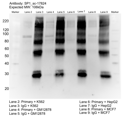
- Caption
- Whole cell lysates of K562, MCF7, GM12878 and HepG2 were immunoprecipitated using the primary antibody (Santa Cruz Biotechnology; sc-636). The IP fraction was separated on a 12% acrylamide gel with the Bio-Rad PROTEAN II xi system. After separation, the samples were transferred to a nitrocellulose membrane with an Invitrogen iBlot system. The membrane was probed with the primary antibody (same as that used for IP) and a secondary HRP-conjugated antibody. The resulting bands were visualized with SuperSignal West Femto Solution (Thermo Scientific). Protein Marker (PM) is labeled in kDa. The approximate size of SP1 is ~106 kDa.
- Reviewer comment
- Band of interest is not 50% of overall signal in lane
- Submitted by
- Mark Mackiewicz
- Lab
- Richard Myers, HAIB
- Grant
- U54HG006998
- Download
- SP1_sc-17824_092916_L.png