ENCAB000AKH
Antibody against Homo sapiens RBBP5
Homo sapiens
K562, HepG2, GM12878, SK-N-SH
characterized to standards
Homo sapiens
any cell type or tissue
partially characterized
Homo sapiens
A549, H1
not characterized to standards
- Status
- released
- Source (vendor)
- Bethyl Labs
- Product ID
- A300-109A
- Lot ID
- 2
- Characterized targets
- RBBP5 (Homo sapiens)
- Host
- rabbit
- Clonality
- polyclonal
- Purification
- affinity
- Aliases
- bradley-bernstein:PchAb 153-V
- External resources
Characterizations
RBBP5 (Homo sapiens)
Method: ChIP-string comparison
not reviewed
- Caption
- Antibody was validated by use of correlation analysis of ChIP-String and ChIP-Seq data
- Submitted by
- Bradley Bernstein
- Lab
- Bradley Bernstein, Broad
- Grant
- U54HG004570
- Download
- human_RBBP5_validation_Bernstein.pdf
RBBP5 (Homo sapiens)
Method: immunoblot
not reviewed
- Submitted by
- Bradley Bernstein
- Lab
- Bradley Bernstein, Broad
- Grant
- U54HG004570
- Download
- human_RBBP5_validation_Bernstein.pdf
RBBP5 (Homo sapiens)
Method: ChIP-seq comparison
compliant
- Submitter comment
- SAV10
- Submitted by
- Nina Farrell
- Lab
- Bradley Bernstein, Broad
- Grant
- U54HG006991
- Download
- RbBP5 PchAb 153-V SAV.pdf
RBBP5 (Homo sapiens)
K562K562
Method: immunoprecipitation
not compliant
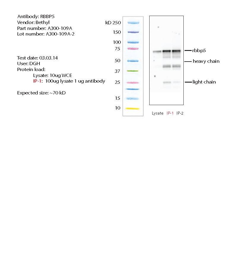
- Caption
- K562 whole cell lysate was immunoprecipitated using primary antibody, and the IP fraction along side a whole cell lysate were loaded on a CriterionXT gel and separated. After separation, the samples were transferred using a wet transfer. Blotting with primary (same as that used for IP) and secondary HRP-conjugated antibodies was performed. Band of expected size visualized representing strongest signal in the lane.
- Reviewer comment
- no IgG shown
- Submitted by
- Noam Shoresh
- Lab
- Bradley Bernstein, Broad
- Grant
- U54HG006991
- Download
- rbbp5_bethyl_A300-109A-2_IP.png
RBBP5 (Homo sapiens)
A549H1
Method: immunoblot
not compliant
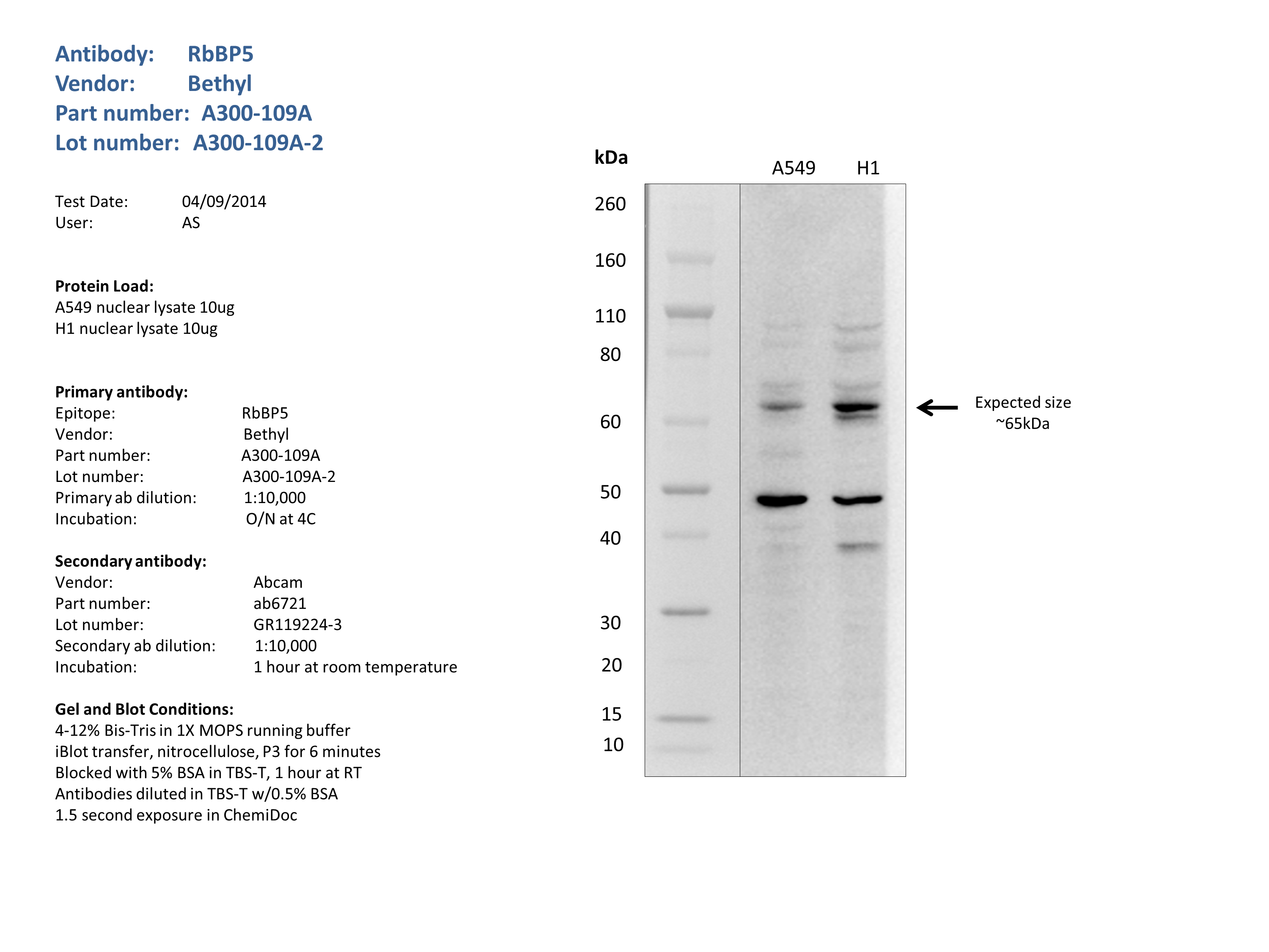
- Caption
- Nuclear lysates from A549 (10ug) and H1 (10ug) were loaded into a 4-12% Bis-Tris gel in 1X MOPS running buffer. After separation, the samples were transferred to a nitrocellulose membrane using iblot. Membrane was blocked for an hour in room temperature, with 5% BSA in TBS-T and blotted with primary antibody in the appropriate concentration over night at 4c. Membrane was washed and blotted with secondary HRP-conjugated antibody. Detection was made with Optiblot ECL Detect Kit (ab133406) for 2 min. A range of bands were detected outside of the expected size.
- Reviewer comment
- Not 50% of signal for either lane.
- Submitted by
- Noam Shoresh
- Lab
- Bradley Bernstein, Broad
- Grant
- U54HG006991
RBBP5 (Homo sapiens)
K562HepG2GM12878SK-N-SH
Method: immunoblot
compliant
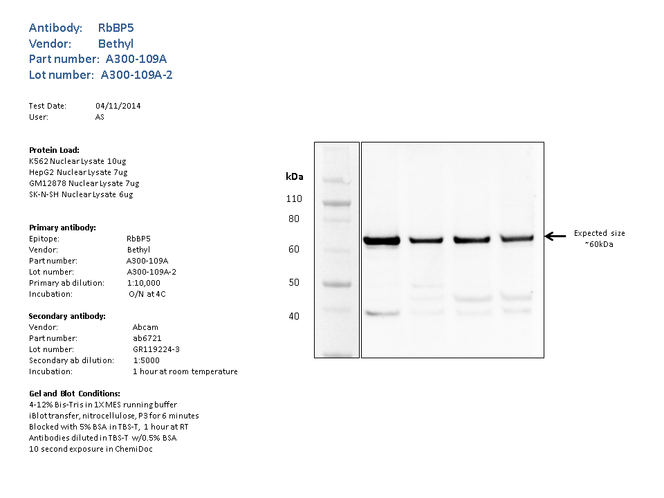
- Caption
- Nuclear lysates from K562 (10ug), HepG2 (7ug), GM12878 (7ug), SK-N-SH (6ug), were loaded into a 4-12% Bis-Tris gel in 1X MES running buffer. After separation, the samples were transferred to a nitrocellulose membrane using the iblot system. Membrane was blocked for an hour in room temperature, with 5% BSA in TBS-T and blotted with primary antibody in the appropriate concentration over night at 4c. Membrane was washed and blotted with secondary HRP-conjugated antibody. Detection was made with Optiblot ECL Detect Kit (ab133406) for 2 min.the strongest band was detected around the expected size (~60kDa).
- Submitted by
- Noam Shoresh
- Lab
- Bradley Bernstein, Broad
- Grant
- U54HG006991