ENCAB000AJK
Antibody against Mus musculus REST, Homo sapiens REST
Homo sapiens
liver, GM23338
characterized to standards with exemption
Mus musculus
any cell type or tissue
partially characterized
Homo sapiens
K562, GM12878
not characterized to standards
- Status
- released
- Source (vendor)
- David Anderson
- Product ID
- NRSF
- Lot ID
- 20070905JV
- Characterized targets
- REST (Mus musculus), REST (Homo sapiens)
- Host
- mouse
- Clonality
- monoclonal
- External resources
Characterizations
REST (Homo sapiens)
Method: motif enrichment
compliant
- Caption
- The motif for target REST is represented by the attached position weight matrix (PWM) derived from files ENCFF574NIK and ENCFF472IUJ. Motif enrichment analysis was done by Dr. Zhizhuo Zhang (Broad Institute, Kellis Lab) using known motifs (http://compbio.mit.edu/encode-motifs/) and previously published ChIP-seq data (http://www.broadinstitute.org/~zzhang/motifpipeline/data/TrainSetInfo.txt). The accept probability score of the given transcription factor was calculated using a Bayesian approach. This analysis also includes three motif enrichment scores, computed by overlapping the motif instances with the given ChIP-seq peak locations. For more information on the underlying statistical methods, please see the attached document. From ENCFF574NIK: Accept probability score: 0.957933586771, Global enrichment Z-score: 7.28890467545, Positional bias Z-score: 13.0430163596, Peak rank bias Z-score: 14.9824152022. From ENCFF472IUJ: Accept probability score: 0.957933586771, Global enrichment Z-score: 6.09615423963, Positional bias Z-score: 12.7323404819, Peak rank bias Z-score: 14.9195663548, Enrichment rank: 1.0.
- Submitted by
- Aditi Narayanan
- Lab
- Richard Myers, HAIB
- Grant
- U54HG006998
- Download
- ENCAB000AJK.pdf
REST (Mus musculus)
Method: immunoprecipitation
not reviewed
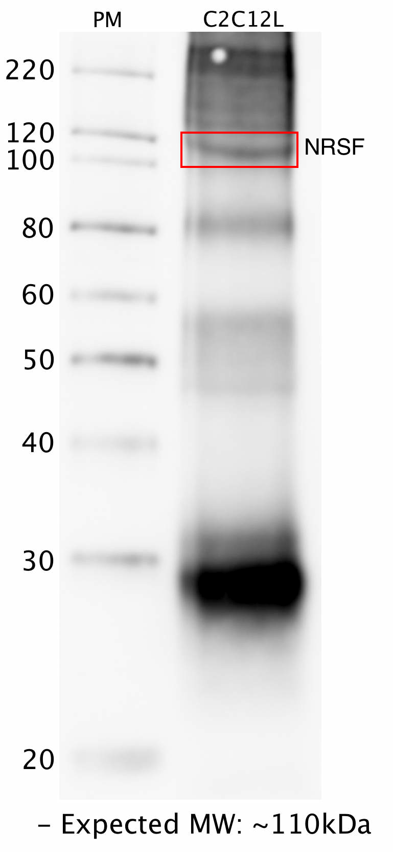
- Caption
- Whole lysate from C2C12 cells was immunoprecipitated with the NRSF (Anderson lab) antibody. The IP fraction was loaded on a 12% acrylamide gel and separated with a Bio-Rad PROTEAN II xi system. After separation, the samples were transferred to a nitrocellulose membrane with an Invitrogen iBlot system. The membrane was then blotted with primary antibody (same as that used for IP) and then a secondary HRP-conjugated antibody. The resulting bands were visualized using SuperSignal West Femto solution (Thermo Scientific). A band of expected size for the NRSF target (~110 kD) was detected, along with contamination from the heavy chain (~50 kD) and light chain (~30kD) of the primary antibody used for IP,
- Submitted by
- Flo Pauli-Behn
- Lab
- Richard Myers, HAIB
- Grant
- U54HG004576
- Download
- HAIB_NRSF_mouse_IPwestern.png
REST (Homo sapiens)
K562GM12878
Method: immunoblot
not compliant
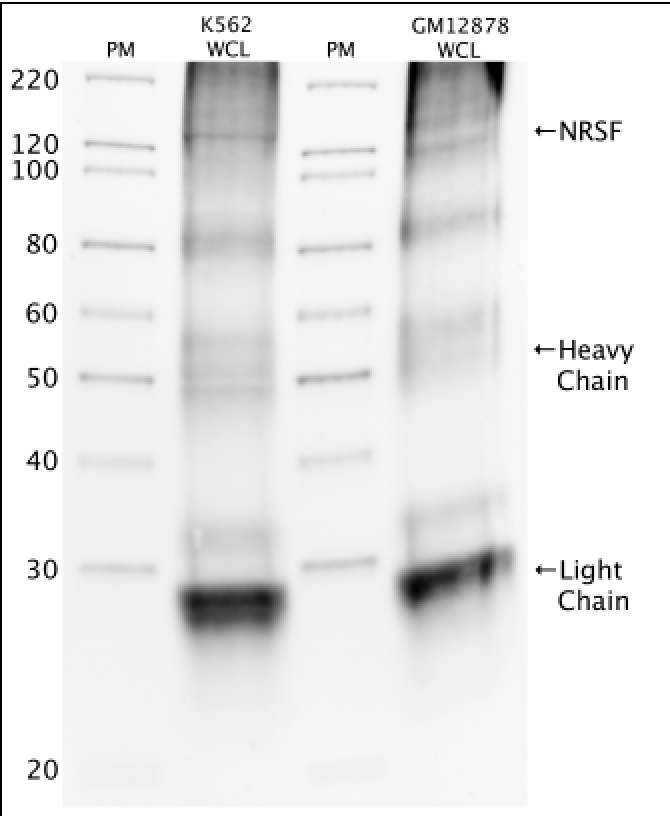
- Caption
- Western blot protocol: Whole cell lysates were immunoprecipitated using primary antibody (Anderson NRSF), and the IP fraction was loaded on a 12% acrylamide gel and separated with a Bio-Rad PROTEAN II xi system. After separation, the samples were transferred to a nitrocellulose membrane with an Invitrogen iBlot system. Blotting with primary (same as that used for IP) and secondary HRP-conjugated antibodies was performed on an Invitrogen BenchPro 4100 system. Visualization was achieved using SuperSignal West Femto solution (Thermo Scientific). Results: Band of expected size visualized, representing strongest signal in the lane. Figure legend: IP-western with NRSF in WCL (whole cell lysates) of K562 and GM12878; PM=protein marker. NRSF band is indicated.
- Reviewer comment
- Band of interest is not 50% of overall signal in lane
- Submitted by
- Richard Myers
- Lab
- Richard Myers, HAIB
- Grant
- U54HG004576
REST (Homo sapiens)
Method: motif enrichment
not reviewed
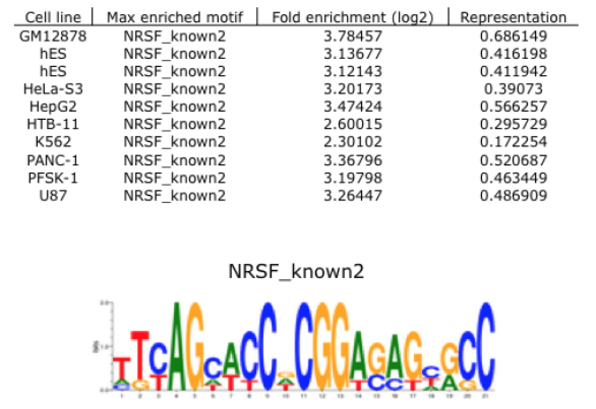
- Caption
- Motif enrichment validation protocol: Motif enrichment validation was performed by Pouya Kheradpour. Methods and results are available at http://www.broadinstitute.org/~pouyak/encode-motif-disc/. Briefly, motif identification was performed on 100 bp peaks (50 bp on either side of peak summit), passing IDR between replicate experiments at < .01. Identified motifs were matched to known motifs for the tested TF (correlation > .75). ENCODE standards call for > 4-fold enrichment and > 10% motif representation in order to consider the identified motif validated. These standards are met and represented in the attached file, which includes cell line tested, name of maximally enriched motif, fold-enrichment value (as log2), and representation percentage. Additionally, known motif consensus sequences which were confirmed experimentally are displayed.
- Submitted by
- Richard Myers
- Lab
- Richard Myers, HAIB
- Grant
- U54HG004576
REST (Homo sapiens)
GM23338
Method: immunoblot
exempt from standards
- Caption
- The ENCODE Binding Working Group finds for some valuable biosamples that recreating a primary on well characterized antibodies is not cost effective. Therefore, they allow exemption from standards for these samples.
- Submitter comment
- The lab is asking for an exemption forIPSCs due to the lack of resources to make a primary characterization for them
- Reviewer comment
- The Antibody Review Board exempted this on 02-19-18. The data within this experiment were released despite a partially characterized antibody in this cell type because the antibody has been characterized to standards in other tissues.
- Submitted by
- Richard Myers
- Lab
- Richard Myers, HAIB
- Grant
- U54HG006998
- Download
- No_biosample.png
REST (Homo sapiens)
liver
Method: immunoblot
exempt from standards
- Caption
- The ENCODE Binding Working Group finds for some valuable tissues that recreating a primary on well characterized antibodies is not cost effective. Therefore, they allow exemption from standards for these tissues.
- Submitter comment
- The lab is asking for an exemption for liver cells due to the lack of resource to make a primary characterization for them
- Reviewer comment
- Exempted by the Feb 29, 2016 antibody review panel
- Submitted by
- Richard Myers
- Lab
- Richard Myers, HAIB
- Grant
- U54HG006998
- Download
- No_tissue.png