ENCAB000AIW
Antibody against Mus musculus MYOG
Mus musculus
C2C12
not characterized to standards
Homo sapiens
LHCN-M2
not characterized to standards
- Status
- released
- Source (vendor)
- Santa Cruz Biotech
- Product ID
- sc-12732
- Lot ID
- unknown
- Characterized targets
- MYOG (Mus musculus)
- Host
- mouse
- Clonality
- monoclonal
- Antigen description
- Epitope mapping within amino acids 138-158 of myogenin of rat origin.
- External resources
Characterizations
MYOG (Mus musculus)
Method: immunoprecipitation followed by mass spectrometry
compliant
- Caption
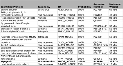
- LHCN whole cell lysate was immunoprecipitated with the myogenin (Santa Cruz Biotech, sc-12732) antibody. The IP fraction was loaded on a 12% acrylamide gel and separated with a Bio-Rad PROTEAN II xi system. The gel was stained with Coomassie Blue in order to visualize marker bands. Gel fragments corresponding to the bands indicated in validation method 1 (IP-western) were excised and sent to the University of Alabama at Birmingham Cancer Center Mass Spectrometry/Proteomics Shared Facility. There the samples were run on an LTQ XL Linear Ion Trap Mass Spectrometer by LC-ESI-MS/MS. Peptides were identified using SEQUEST tandem mass spectra analysis, with probability based matching at p < 0.05. Scaffold software (v4.0.3, protein threshold of 99% minimum, 2 peptides minimum, and protein FDR of 0.1% or less) was then used to identify proteins in the mouse genome that match the peptides identified by SEQUEST. The Scaffold results are listed in the table. The myogenin target was identified along with common contaminants.
- Reviewer comment
- Protein identified in mass spec, but the full table is required to be submitted as stated in the standards document.
- Submitted by
- Flo Pauli-Behn
- Lab
- Richard Myers, HAIB
- Grant
- U54HG004576
- Download
- HAIB_myogenin_mouse_IPmassspec.png
MYOG (Mus musculus)
C2C12LHCN-M2
Method: immunoprecipitation
not compliant
- Caption
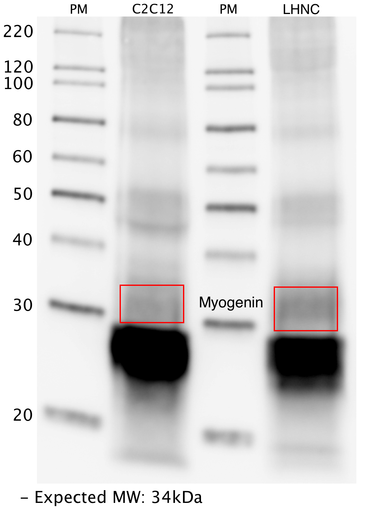
- Whole lysate from C2C12 and LHNC cells were immunoprecipitated with the myogenin (Santa Cruz Biotech, sc-12732) antibody. The IP fraction was loaded on a 12% acrylamide gel and separated with a Bio-Rad PROTEAN II xi system. After separation, the samples were transferred to a nitrocellulose membrane with an Invitrogen iBlot system. The membrane was then blotted with primary antibody (same as that used for IP) and then a secondary HRP-conjugated antibody. The resulting bands were visualized using SuperSignal West Femto solution (Thermo Scientific). A band of expected size for the myogenin target (~34 kD) was detected, along with contamination from the light chain (~28kD) of the primary antibody used for IP.
- Submitter comment
- no control
- Submitted by
- Flo Pauli-Behn
- Lab
- Richard Myers, HAIB
- Grant
- U54HG004576