ENCAB000AIJ
Antibody against Homo sapiens MAFK, Mus musculus MAFK
Homo sapiens
HepG2
characterized to standards
Homo sapiens
any cell type or tissue
partially characterized
Mus musculus
any cell type or tissue
partially characterized
Homo sapiens
GM12878, K562, HeLa-S3
not characterized to standards
- Status
- released
- Source (vendor)
- Abcam
- Product ID
- ab50322
- Lot ID
- 904274
- Characterized targets
- MAFK (Homo sapiens), MAFK (Mus musculus)
- Host
- rabbit
- Clonality
- polyclonal
- Purification
- other
- Antigen description
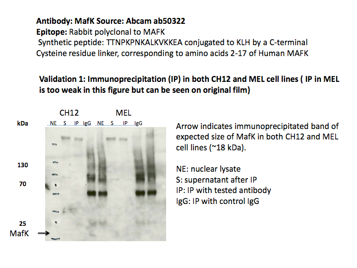
- Raised against synthetic peptide: TTNPKPNKALKVKKEA conjugated to KLH by a C-terminal Cysteine residue linker, corresponding to amino acids 2-17 of Human MAFK.
- Antigen sequence
- TTNPKPNKALKVKKEA
- External resources
Characterizations
MAFK (Homo sapiens)
Method: motif enrichment
compliant
- Caption
- The motif for target MAFK is represented by the attached position weight matrix (PWM) derived from ENCFF0067PZL. Motif enrichment analysis was done by Dr. Zhizhuo Zhang (Broad Institute, Kellis Lab). Accept probability score: 0.99638610008 Global enrichment Z-score: 9.48493269478 Positional bias Z-score: 11.8653123173 Peak rank bias Z-score: 4.79425564585 Enrichment rank: 3.0
- Submitted by
- Kathrina Onate
- Lab
- Michael Snyder, Stanford
- Grant
- U54HG006996
MAFK (Homo sapiens)
Method: motif enrichment
compliant
- Caption
- The motif for target MAFK is represented by the attached position weight matrix (PWM) derived from ENCFF155SYQ. Motif enrichment analysis was done by Dr. Zhizhuo Zhang (Broad Institute, Kellis Lab). Accept probability score: 0.997040296854 Global enrichment Z-score: 9.55502639073 Positional bias Z-score: 12.3654501273 Peak rank bias Z-score: 10.0221573264 Enrichment rank: 2.0
- Submitted by
- Kathrina Onate
- Lab
- Michael Snyder, Stanford
- Grant
- U54HG006996
MAFK (Homo sapiens)
Method: immunoprecipitation
not reviewed
- Caption
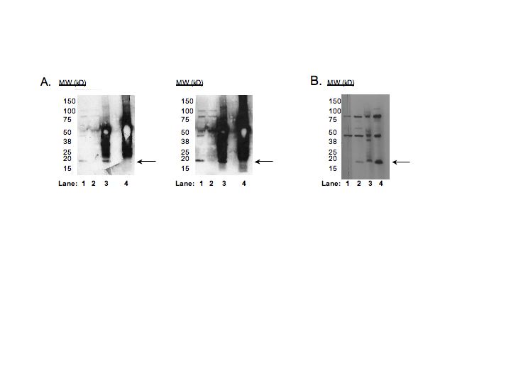
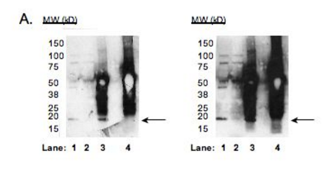
- A: Immunoprecipitation was performed on nuclear lysates from HepG2 cells using antibody ab50322 against MafK/NF-E2 p18. Lane1: Nuclear lysate.Lane 2: Unbound material from immunoprecipitation with ab50322.Lane 3: Bound material from immunoprecipitation with ab50322.Lane 4:Bound material from control immunoprecipitation with rabbit IgG.Arrow indicates band of expected size (18kD) that is highly enriched in the specifically immunoprecipitated fraction.Left panel contains a shorter exposure to emphasize the identity of the expected band while right panel is overexposed to emphasize that all bands other than the expected band are present in the non-immunoprecipitated flowthrough lane (lane 2).Note that ~6x more material is loaded in lanes 3 and 4, which explains the enrichment of the expected product in the immunoprecipitated fraction relative to the input nuclear lysate in lane 1. B:Western blots on nuclear lysates from cell lines GM12878 (Lane1), K562 (Lane2), HeLaS3 (Lane3), and HepG2 (Lane4). COMMENT:While we observe multiple bands in the Western blots for all cell lines, we see that only the expected band is immunoprecipitated specifically and efficiently.Furthermore, because we have performed ChIP-Seq with a second antibody recognizing an independent epitope and our results show a very high level of agreement (see second validation), we feel this antibody isof high quality.
- Submitted by
- Michael Snyder
- Lab
- Michael Snyder, Stanford
- Grant
- U54HG004558
MAFK (Homo sapiens)
Method: ChIP-seq comparison

not reviewed
- Caption
- Validation 2:ChIP-Seq with alternate antibody: Santa Cruz sc-477 Epitope: Rabbit polyclonal, epitope maps to the C-terminus of MafK/NF-E2 p18 of mouse origin Background/Comparison:The small Maf family of proteins consists of three small, highly similar proteins- MafF, MafG, and MafK.An alignment of the three is shown here.Due to the high degree of similarity between these factors, we have used two antibodies to different regions of the MafK protein to cross-validate our ChIP-Seq results.The epitope for sc-477 is indicated in red and the immunogen for ab50322 is indicated in yellow. The lists of peaks called by PeakSeq obtained from the two ENCODE-submitted biological replicate experiments done for each antibody in HepG2 cells were compared to one another.Comparisons were made using standard ENCODE replicate comparison parameters (the top 40% of each list of peaks for a given false discovery rate (Q-value) were compared to the entire list peaks obtained withthe other antibody and the reciprocal comparison was also made).Comparisons were made for Q-values of .01 and .001 and the results are shown in the accompanying table.In each case, the degree of overlap between peak lists exceeds 97%.Thus, these antibodies are considered validated by this criteria.
- Submitted by
- Michael Snyder
- Lab
- Michael Snyder, Stanford
- Grant
- U54HG004558
MAFK (Homo sapiens)
HepG2
Method: immunoprecipitation
compliant
- Caption
- A: Immunoprecipitation was performed on nuclear lysates from HepG2 cells using antibody ab50322 against MafK/NF-E2 p18. Lane1: Nuclear lysate. Lane 2: Unbound material from immunoprecipitation with ab50322. Lane 3: Bound material from immunoprecipitation with ab50322. Lane 4:Bound material from control immunoprecipitation with rabbit IgG. Arrow indicates band of expected size (18kD) that is highly enriched in the specifically immunoprecipitated fraction. Left panel contains a shorter exposure to emphasize the identity of the expected band while right panel is overexposed to emphasize that all bands other than the expected band are present in the non-immunoprecipitated flowthrough lane (lane 2). Note that ~6x more material is loaded in lanes 3 and 4, which explains the enrichment of the expected product in the immunoprecipitated fraction relative to the input nuclear lysate in lane 1.
- Submitted by
- Kathrina Onate
- Lab
- Michael Snyder, Stanford
- Grant
- U54HG004558
- Download
- IP Snyder AIJ.png
MAFK (Mus musculus)
Method: immunoprecipitation
not reviewed
- Caption
- The immunogen used to generate this antibody is identical in mouse and human and the proteins share 98% identity over their full lengths. Only one major band (indicated by arrow) is observed in an immunoblot analysis of material immunoprecipitated by ab50322 from CH12 cells and is absent in material immunoprecipitated with control IgG. This band, indicated by arrow, has a mobility consistent with the expected size of MafK (~18kD).Additional bands observed by immunoblot with nuclear lysate are not efficiently immunoprecipitated. Similar results are obtained in MEL cells, although the immunoblot signal is weaker. This antibody has been previously validated by immunoblot in human cell lines K562, HeLaS3, GM12878, and HepG2, by immunoprecipitation in HepG2 cells, and by ChIPSeq using two independent antibodies in HepG2 cells and this validation information has been submitted. In combination with the previously submitted data, ab50322 meets the ENCODE criteria for validation in murine cell lines CH12 and MEL.
- Submitted by
- Michael Snyder
- Lab
- Michael Snyder, Stanford
- Grant
- RC2HG005602
MAFK (Homo sapiens)
GM12878K562HeLa-S3HepG2
Method: immunoblot
not compliant
- Caption
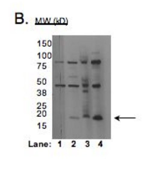
- B: Western blots on nuclear lysates from cell lines GM12878 (Lane1), K562 (Lane2), HeLaS3 (Lane3), and HepG2 (Lane4). COMMENT: While we observe multiple bands in the Western blots for all cell lines, we see that only the expected band is immunoprecipitated specifically and efficiently. Furthermore, because we have performed ChIP-Seq with a second antibody recognizing an independent epitope and our results show a very high level of agreement (see second validation), we feel this antibody is of high quality.
- Reviewer comment
- Multiple bands and band of expected size does not have the strongest signal.
- Submitted by
- Kathrina Onate
- Lab
- Michael Snyder, Stanford
- Grant
- U54HG004558
- Download
- WB Snyder AIJ.png