ENCAB000AGR
Antibody against Homo sapiens GABPA
Homo sapiens
K562
characterized to standards
Homo sapiens
HeLa-S3, A-431, NIH3T3, liver, GM12878, HepG2, MCF-7
characterized to standards with exemption
- Status
- released
- Source (vendor)
- Santa Cruz Biotech
- Product ID
- sc-28312
- Lot ID
- F1804
- Characterized targets
- GABPA (Homo sapiens)
- Host
- mouse
- Clonality
- monoclonal
- Purification
- crude
- Isotype
- IgG1
- Antigen description
- raised against amino acids 1-180 of GABP-α of human origin
- Antigen sequence
- MTKREAEELIEIEIDGTEKAECTEESIVEQTYAPAECVSQAIDINEPIGNLKKLLEPRLQCSLDAHEICLQDIQLDPERSLFDQGVKTDGTVQLSVQVISYQGIEPKLNILEIVKPADTVEVVIDPDAHHAESEAHLVEEAQVITLDGTKHITTISDETSEQVTRWAAALEGYRKEQER
- External resources
Characterizations
GABPA (Homo sapiens)
Method: immunoprecipitation followed by mass spectrometry
compliant
- Caption
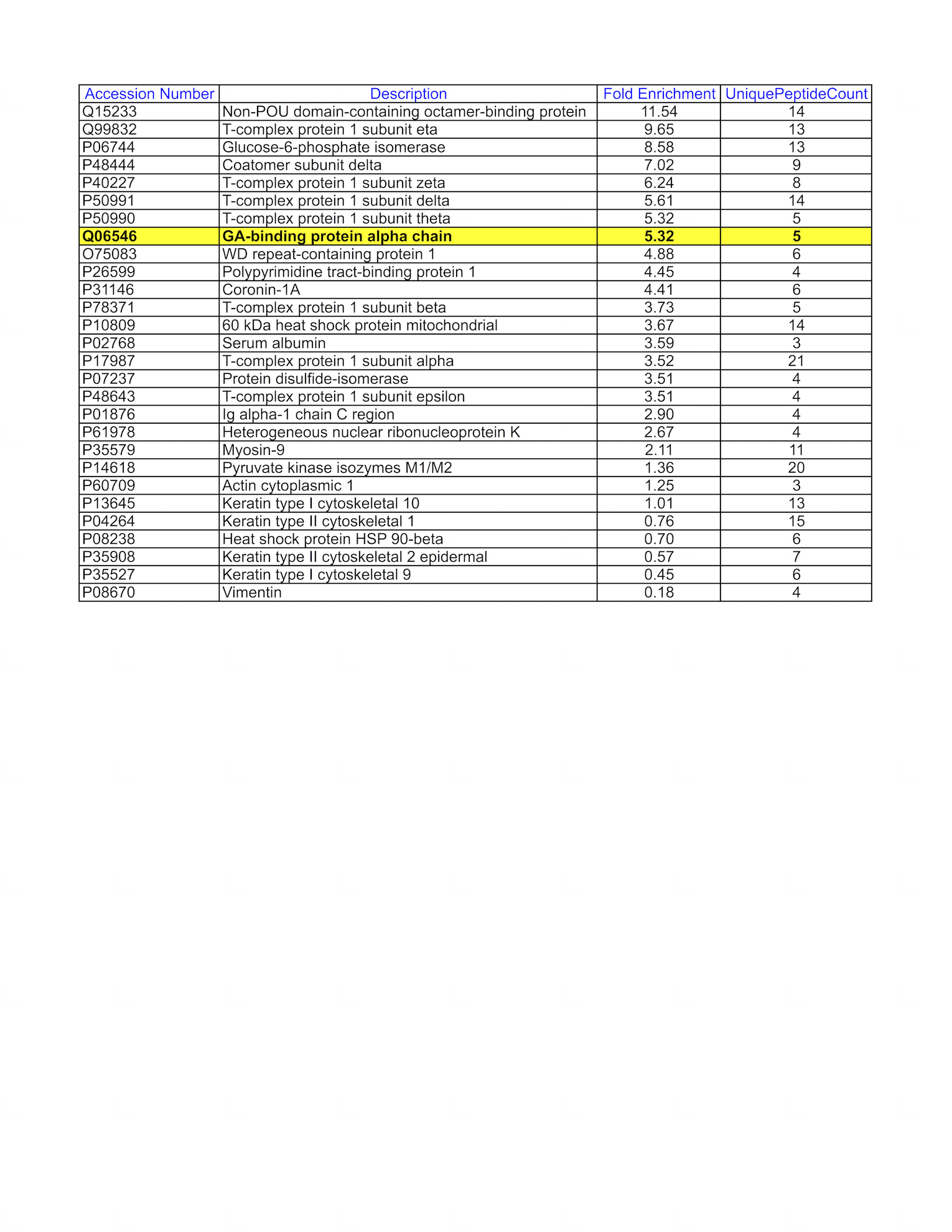
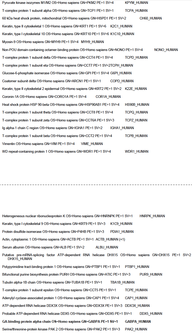
- GM12878 whole cell lysate was immunoprecipitated using the primary antibody (Santa Cruz Biotechnology; sc-28312). The IP fraction was loaded on a 12% Bio-Rad TGX gel and separated with the Bio-Rad Tetra Cell system. A gel fragment corresponding to a band on a Coomassie Blue stained gel image was excised and sent to the University of Alabama at Birmingham Cancer Center Mass Spectrometry/Proteomics Shared Facility. Analysis of gel fragment from GM12878: The sample was analyzed on a LTQ XL Linear Ion Trap Mass Spectrometer by LC-ESI-MS/MS. Peptides were identified using SEQUEST tandem mass spectral analysis with probability based matching at p < 0.05. SEQUEST results were reported with ProteinProphet protXML Viewer (TPP v4.4 JETSTREAM) and filtered for a minimum probability of 0.9. All protein hits that met these criteria were reported, including common contaminants. Fold enrichment for each protein reported was determined using a custom script based on the FC-B score calculation from the reference Mellacheruvu et al., 2013. The CRAPome: a contaminant repository for affinity purification mass spectrometry data. Nat. Methods. 10(8):730-736. Doi:10.1038/nmeth.2557. The target protein, GABPA, was identified as the 8th ranked enriched protein and the 1st ranked transcription factor based on IP-Mass Spectrometry.
- Submitted by
- Mark Mackiewicz
- Lab
- Richard Myers, HAIB
- Grant
- U54HG006998
- Download
- GABPA_IP-MS.png
GABPA (Homo sapiens)
K562GM12878HepG2MCF-7
Method: immunoprecipitation
exempt from standards
- Caption
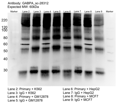
- Whole cell lysates of K562, MCF7, and HepG2 were immunoprecipitated using the primary antibody (Santa Cruz Biotechnology; sc-28312). The IP fraction was separated on a 12% acrylamide gel with the Bio-Rad PROTEAN II xi system. After separation, the samples were transferred to a nitrocellulose membrane with an Invitrogen iBlot system. The membrane was probed with the primary antibody (same as that used for IP) and a secondary HRP-conjugated antibody. The resulting bands were visualized with SuperSignal West Femto Solution (Thermo Scientific). Protein Marker (PM) is labeled in kDa. The approximate size of GABPA is ~60 kDa.
- Submitter comment
- --
- Reviewer comment
- Band of interest is not 50% of overall signal in lane. Another stronger band is closer to the size we would expect (50 kDa). Rescued by mass spec
- Submitted by
- Mark Mackiewicz
- Lab
- Richard Myers, HAIB
- Grant
- U54HG004576
- Download
- GABPA_sc-28312_092916_L.png
GABPA (Homo sapiens)
liver
Method: immunoblot
exempt from standards
- Caption
- The ENCODE Binding Working Group finds for some valuable tissues that recreating a primary on well characterized antibodies is not cost effective. Therefore, they allow exemption from standards for these tissues.
- Submitter comment
- The lab is asking for an exemption for liver cells due to the lack of resource to make a primary characterization for them
- Reviewer comment
- Exempted by the Feb 29, 2016 antibody review panel
- Submitted by
- Richard Myers
- Lab
- Richard Myers, HAIB
- Grant
- U54HG006998
- Download
- No_tissue.png
GABPA (Homo sapiens)
K562
Method: immunoblot
compliant
- Caption
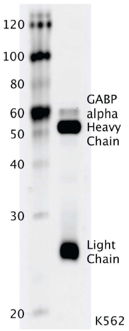
- Whole cell lysates were immunoprecipitated using primary antibody, and the IP fraction was loaded on a 12% acrylamide gel and separated with a Bio-Rad PROTEAN II xi system. After separation, the samples were transferred to a nitrocellulose membrane using a Bio-Rad Trans-Blot Electrophoretic Transfer system. Standard western blot protocol was used to probe the membrane with the primary antibody (same antibody as used for IP), and an HRP-conjugated secondary antibody and SuperSignal West Femto solution (Thermo Scientific) were used to detect the immunoprecipitated proteins. Figure Legend: GABPA immunoblot: IP-western with sc-28312 GABP-α antibody in whole cell lysate of K562. Heavy chain and light chain of IgG are indicated, and GABPA band is indicated at ~60 kDa.
- Submitted by
- Marcus Ho
- Lab
- Richard Myers, HAIB
- Grant
- U54HG004576
GABPA (Homo sapiens)
Method: immunoprecipitation followed by mass spectrometry
not compliant
- Caption
- ENCODE data standards recognizes various methodologies for secondary validation of antibodies. Among these methodologies is immunoprecipitation followed by mass spectrometry analysis. Briefly, GM12878 whole cell lysates were immunoprecipitated using primary antibody, and the IP fraction was loaded on a 12% acrylamide gel and separated with a Bio-Rad PROTEAN II xi system. Gel was stained with Coomasie Blue in order to visualize marker bands. A gel fragment corresponding to the band indicated above in the western blot image was excised and sent to the University of Alabama at Birmingham Cancer Center Mass Spectrometry/Proteomics Shared Facility. There the sample was run on an LTQ XL Linear Ion Trap Mass Spectrometer with alternating collision-induced dissociation and electron-transfer dissociation. Peptides were identified using MASCOT (Matrix Science), with probability based matching at p < 0.05. Subsequent analysis was performed in Scaffold (Proteome Software, Inc.) at 0.0% protein FDR and 0.0% peptide FDR. As per ENCODE data standards, all Scaffold results are listed below, including common contaminants. Target protein is highlighted in bold font.
- Reviewer comment
- (VENKAT)- "need to add caption. should the "fold enrichment doc - /documents/3bcdcea7-f0ed-4a74-b94c-4e5eeb1ec7a0/ " be added?"
- Submitted by
- Richard Myers
- Lab
- Richard Myers, HAIB
- Grant
- U54HG004576
GABPA (Homo sapiens)
HeLa-S3A-431NIH3T3
Method: immunoprecipitation
exempt from standards
- Caption
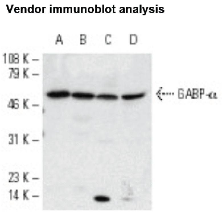
- For an antibody to meet ENCODE validation standards, a single band of the predicted size, or a band of no less than half the total signal, must be detected in a lane on a Western blot. Figure Legend: Western blot analysis of GABP-α expression in HeLa (A), A- 431 (B), NIH/3T3 (C) and 3611-RF (D) nuclear extracts. Expected size: ~51 kDa
- Submitter comment
- --
- Reviewer comment
- The antibody review panel has decided that antibodies that pass ENCODE2 standards, and are only missing their IgG controls for ENCODE3 standards, will be passed via exemption.
- Submitted by
- Richard Myers
- Lab
- Richard Myers, HAIB
- Grant
- U54HG004576