ENCAB000AFC
Antibody against Homo sapiens CEBPD
Homo sapiens
HepG2, HeLa-S3, liver, K562, MCF-7
not characterized to standards
- Status
- released
- Source (vendor)
- Santa Cruz Biotech
- Product ID
- sc-636
- Lot ID
- C1010
- Characterized targets
- CEBPD (Homo sapiens)
- Host
- rabbit
- Clonality
- polyclonal
- Isotype
- IgG
- Antigen description
- Epitope mapping at the C-terminus of CEBPD of human origin.
- External resources
Characterizations
CEBPD (Homo sapiens)
K562HepG2MCF-7
Method: immunoprecipitation
not compliant
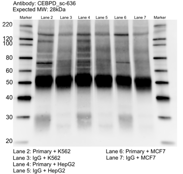
- Caption
- Whole cell lysates of K562, MCF7, and HepG2 were immunoprecipitated using the primary antibody (Santa Cruz Biotechnology; sc-636). The IP fraction was separated on a 12% acrylamide gel with the Bio-Rad PROTEAN II xi system. After separation, the samples were transferred to a nitrocellulose membrane with an Invitrogen iBlot system. The membrane was probed with the primary antibody (same as that used for IP) and a secondary HRP-conjugated antibody. The resulting bands were visualized with SuperSignal West Femto Solution (Thermo Scientific). Protein Marker (PM) is labeled in kDa. The approximate size of CEBPD is ~28 kDa.
- Reviewer comment
- Band of interest is not 50% of overall signal in lane
- Submitted by
- Mark Mackiewicz
- Lab
- Richard Myers, HAIB
- Grant
- U54HG006998
- Download
- CEBPD_sc-636_092916_L.png
CEBPD (Homo sapiens)
Method: immunoprecipitation followed by mass spectrometry
not compliant
- Caption
- IP followed by mass spectrometry: Briefly, HeLa whole cell lysates were immunoprecipitated using primary antibody, and the IP fraction was loaded on a 12% acrylamide gel and separated with a Bio-Rad PROTEAN II xi system. Gel was stained with Coomassie Blue in order to visualize marker bands. Gel fragments corresponding to the bands indicated above in the western blot image were excised and sent to the University of Alabama at Birmingham Cancer Center Mass Spectrometry/Proteomics Shared Facility. There the samples were run on an LTQ XL Linear Ion Trap Mass Spectrometer by LC-ESI-MS/MS. Peptides were identified using SEQUEST tandem mass spectra analysis, with probability based matching at p < 0.05. As per ENCODE data standards, all SEQUEST results are attached (ENCODE_HAIB_CEBPD_(SC-636)_07192011_MassSpec.pdf), including common contaminants. Target protein is listed as hit 28 in lower (~35 kDa) band and not detected in upper (~120 + kDa) bands. No proteins with known DNA-binding activity were detected in the upper bands.
- Reviewer comment
- Not within top 20 factors ranked by fold enrichment. Proper document of results not provided.
- Submitted by
- Richard Myers
- Lab
- Richard Myers, HAIB
- Grant
- U54HG004576
CEBPD (Homo sapiens)
HepG2HeLa-S3
Method: immunoblot
not compliant
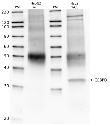
- Caption
- Western blot protocol: Whole cell lysate was immunoprecipitated using primary antibody, and the IP fraction was loaded on a 12% acrylamide gel and separated with a Bio-Rad PROTEAN II xi system. After separation, the samples were transferred to a nitrocellulose membrane with an Invitrogen iBlot system. Blotting with primary (same as that used for IP) and secondary HRP-conjugated antibodies was performed on an Invitrogen BenchPro 4100 system. Visualization was achieved using SuperSignal West Femto solution (Thermo Scientific). Results: Band of expected size visualized, representing strongest signal in the lane. Upper bands also visualized at ~120 kD. Bands at ~35 kD and ~120+ kD tested by IP-mass spec. Only the band at ~35 kD was positive for CEBPD. Figure legend: IP-western with sc-636 in whole cell lysate (WCL) of HepG2 and HeLa; PM=protein marker. CEBPD bands are indicated. Expected size: ~28.5 kDa.
- Reviewer comment
- HepG2: No band visible in the expected size range. HeLa-S3: Band is not at least 50% of overall lane signal
- Submitted by
- Richard Myers
- Lab
- Richard Myers, HAIB
- Grant
- U54HG004576
CEBPD (Homo sapiens)
liver
Method: immunoblot
not compliant
- Caption
- The ENCODE Binding Working Group finds for some valuable tissues that recreating a primary on well characterized antibodies is not cost effective. Therefore, they allow exemption from standards for these tissues.
- Submitter comment
- The lab is asking for an exemption for liver cells due to the lack of resource to make a primary characterization for them.
- Reviewer comment
- Not compliant due to a lack of at least two compliant characterizations in other tissue/cell types.
- Submitted by
- Richard Myers
- Lab
- Richard Myers, HAIB
- Grant
- U54HG006998
- Download
- No_tissue.png