ENCAB000AFB
Antibody against Mus musculus CEBPB, Homo sapiens CEBPB
Homo sapiens
K562, GM12878, HeLa-S3, HepG2
characterized to standards
Mus musculus
any cell type or tissue
partially characterized
Homo sapiens
any cell type or tissue
partially characterized
- Status
- released
- Source (vendor)
- Santa Cruz Biotech
- Product ID
- sc-150
- Lot ID
- I1010
- Characterized targets
- CEBPB (Mus musculus), CEBPB (Homo sapiens)
- Host
- rabbit
- Clonality
- polyclonal
- Isotype
- IgG
- Antigen description
- Epitope mapping at the C-terminus of C/EBP-beta of rat origin
- External resources
Characterizations
CEBPB (Homo sapiens)
Method: motif enrichment
compliant
- Caption
- The motif for target CEBPB is represented by the attached position weight matrix (PWM) derived from ENCFF340TLG. Motif enrichment analysis was done by Dr. Zhizhuo Zhang (Broad Institute, Kellis Lab). Accept probability score: 0.915852871039 Global enrichment Z-score: 9.21272645299 Positional bias Z-score: 8.41147333647 Peak rank bias Z-score: 3.45566069931 Enrichment rank: 1.0
- Submitted by
- Kathrina Onate
- Lab
- Michael Snyder, Stanford
- Grant
- U54HG006996
CEBPB (Mus musculus)
Method: immunoprecipitation
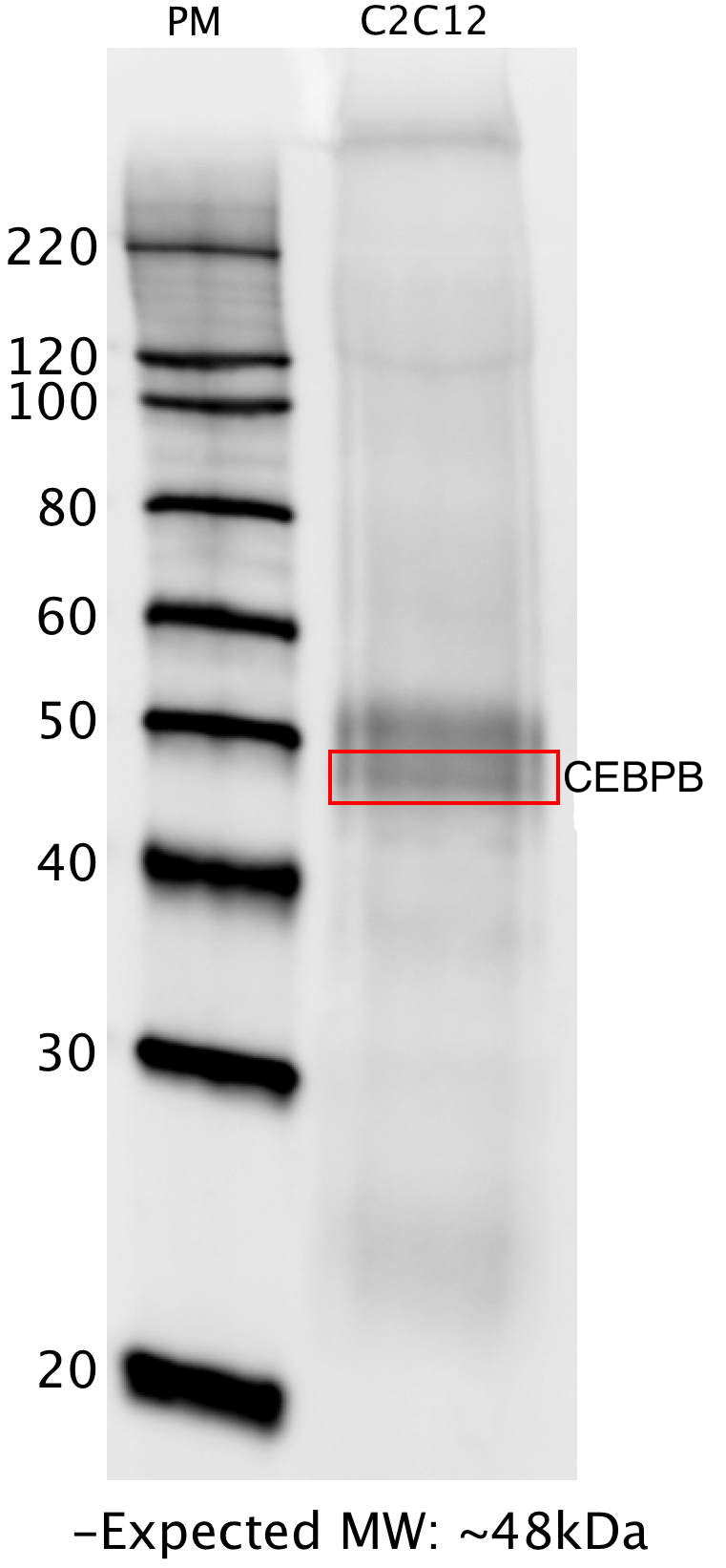
not reviewed
- Caption
- Whole lysate from C2C12 cells was immunoprecipitated with the CEBP (sc-150) antibody. The IP fraction was loaded on a 12% acrylamide gel and separated with a Bio-Rad PROTEAN II xi system. After separation, the samples were transferred to a nitrocellulose membrane with an Invitrogen iBlot system. The membrane was then blotted with primary antibody (same as that used for IP) and then a secondary HRP-conjugated antibody. The resulting bands were visualized using SuperSignal West Femto solution (Thermo Scientific). A band of expected size for the CEBP target (~48 kD) was detected, along with possible contamination from the heavy chain of the primary antibody used for IP (~50 kD).
- Submitted by
- Flo Pauli-Behn
- Lab
- Richard Myers, HAIB
- Grant
- U54HG004576
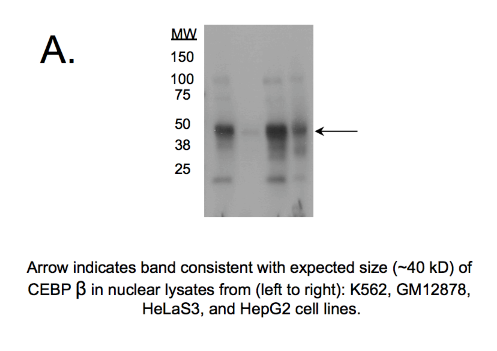
CEBPB (Homo sapiens)
Method: immunoblot
not reviewed
- Caption
- Western blot protocol: Whole cell lysate was immunoprecipitated using primary antibody, and the IP fraction was loaded on a 12% acrylamide gel and separated with a Bio-Rad PROTEAN II xi system. After separation, the samples were transferred to a nitrocellulose membrane with an Invitrogen iBlot system. Blotting with primary (same as that used for IP) and secondary HRP-conjugated antibodies was performed on an Invitrogen BenchPro 4100 system. Visualization was achieved using SuperSignal West Femto solution (Thermo Scientific). Results: Band of expected size visualized, representing strongest signal in the lane. Band was tested by IP-mass spec. Figure legend: IP-western with sc-150 in whole cell lysate (WCL) of K562, HepG2 and HeLa cells; PM=protein marker. CEBPB band is indicated.
- Submitted by
- Richard Myers
- Lab
- Richard Myers, HAIB
- Grant
- U54HG004576
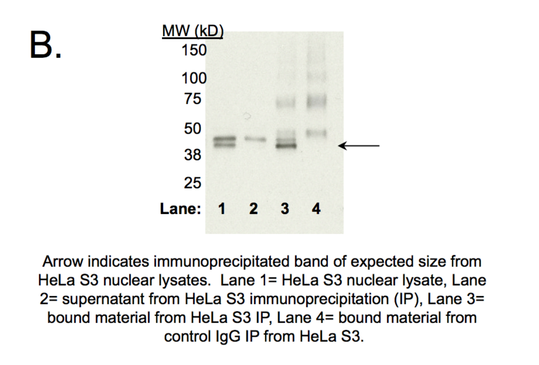
CEBPB (Homo sapiens)
Method: immunoprecipitation followed by mass spectrometry
not reviewed
- Caption
- IP followed by mass spectrometry: Briefly, K562 whole cell lysates were immunoprecipitated using primary antibody, and the IP fraction was loaded on a 12% acrylamide gel and separated with a Bio-Rad PROTEAN II xi system. Gel was stained with Coomassie Blue in order to visualize marker bands. Gel fragments corresponding to the bands indicated above in the western blot image were excised and sent to the University of Alabama at Birmingham Cancer Center Mass Spectrometry/Proteomics Shared Facility. There the samples were run on an LTQ XL Linear Ion Trap Mass Spectrometer by LC-ESI-MS/MS. Peptides were identified using SEQUEST tandem mass spectra analysis, with probability based matching at p < 0.05. As per ENCODE data standards, all SEQUEST results are attached (ENCODE_HAIB_CEBPB_(SC-150)_07192011_MassSpec.pdf), including common contaminants. Target protein is listed as hit 19a in the ~48 kD band.
- Submitted by
- Richard Myers
- Lab
- Richard Myers, HAIB
- Grant
- U54HG004576
CEBPB (Homo sapiens)
Method: immunoprecipitation
not reviewed
- Submitted by
- Michael Snyder
- Lab
- Michael Snyder, Stanford
- Grant
- U54HG004558
- Download
- human_CEBPbeta_validation_Snyder.pdf
CEBPB (Homo sapiens)
Method: motif enrichment
not reviewed
- Submitted by
- Michael Snyder
- Lab
- Michael Snyder, Stanford
- Grant
- U54HG004558
- Download
- human_CEBPbeta_validation_Snyder.pdf
CEBPB (Homo sapiens)
HeLa-S3
Method: immunoprecipitation
compliant
- Submitted by
- Kathrina Onate
- Lab
- Michael Snyder, Stanford
- Grant
- U54HG004558
- Download
- IP Snyder AFB.png
CEBPB (Homo sapiens)
GM12878
Method: immunoprecipitation
compliant
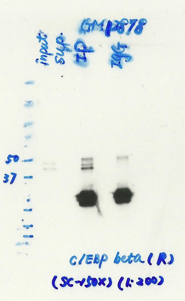
- Caption
- Immunoprecipitation was performed on nuclear extracts from the cell line: GM12878, using the antibody sc-150x. The blot shows western blot analysis of input, flowthrough, immunoprecipitate and mock immunoprecipitate using IgG.Molecular Weight: 36.106. There is an isoform ~33 kD (P17676-2) that should account for other similarly sized band seen in the picture.
- Submitted by
- Nathaniel Watson
- Lab
- Michael Snyder, Stanford
- Grant
- U54HG006996
- Download
- Scan_20160321.jpg
CEBPB (Homo sapiens)
K562GM12878HeLa-S3HepG2
Method: immunoblot
compliant
- Reviewer comment
- Lane 2 (GM12878) does not seem to correspond >50% of all bands
- Submitted by
- Kathrina Onate
- Lab
- Michael Snyder, Stanford
- Grant
- U54HG004558
- Download
- WB Snyder AFB.png