ENCAB000AEK
Antibody against Homo sapiens BHLHE40, Mus musculus BHLHE40
Homo sapiens
K562, IMR-90, GM12878, HeLa-S3, HepG2, HEK293T
characterized to standards
Homo sapiens
any cell type or tissue
partially characterized
Mus musculus
any cell type or tissue
partially characterized
- Status
- released
- Source (vendor)
- Novus
- Product ID
- NB100-1800
- Lot ID
- A1
- Characterized targets
- BHLHE40 (Homo sapiens), BHLHE40 (Mus musculus)
- Host
- rabbit
- Clonality
- polyclonal
- Purification
- other
- Antigen description
- maps to a region between residues 350 and the C-terminus (residue 412).
- External resources
Characterizations
BHLHE40 (Homo sapiens)
IMR-90IMR-90
Method: immunoprecipitation
compliant
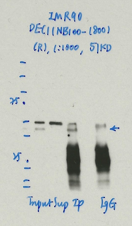
- Caption
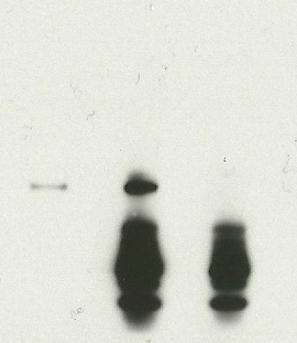
- Immunoprecipitation was performed on nuclear extracts from the cell line: IMR90, using the antibody NB100-1800. The blot shows western blot analysis of input, flowthrough, immunoprecipitate and mock immunoprecipitate using IgG.Molecular Weight: 51.0
- Reviewer comment
- UniProt says the size is ~45.5kD (O14503-1).
- Submitted by
- Nathaniel Watson
- Lab
- Michael Snyder, Stanford
- Grant
- U54HG006996
- Download
- 1128_08_DEC1_NB100-1800.jpg
BHLHE40 (Homo sapiens)
GM12878K562HeLa-S3HepG2
Method: immunoblot
compliant
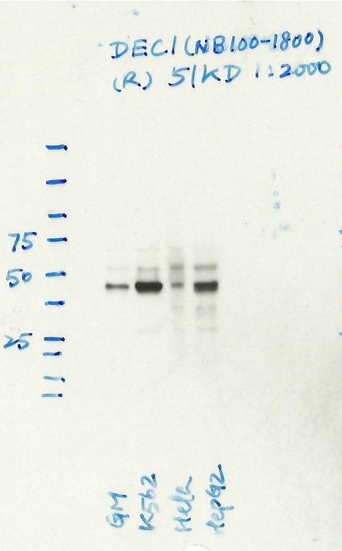
- Caption
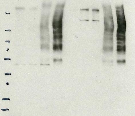
- Western blot analysis of nuclear lysates prepared from multiple cells lines loaded in the order: GM12878, K562, HeLaS3, HepG2 using the antibody NB100-1800. Molecular Weight: 51.0
- Reviewer comment
- Running a bit lower than the expected size...
- Submitted by
- Nathaniel Watson
- Lab
- Michael Snyder, Stanford
- Grant
- U54HG006996
- Download
- BHLHE40(DEC1)-WB-175.jpg
BHLHE40 (Homo sapiens)
K562
Method: immunoprecipitation
compliant
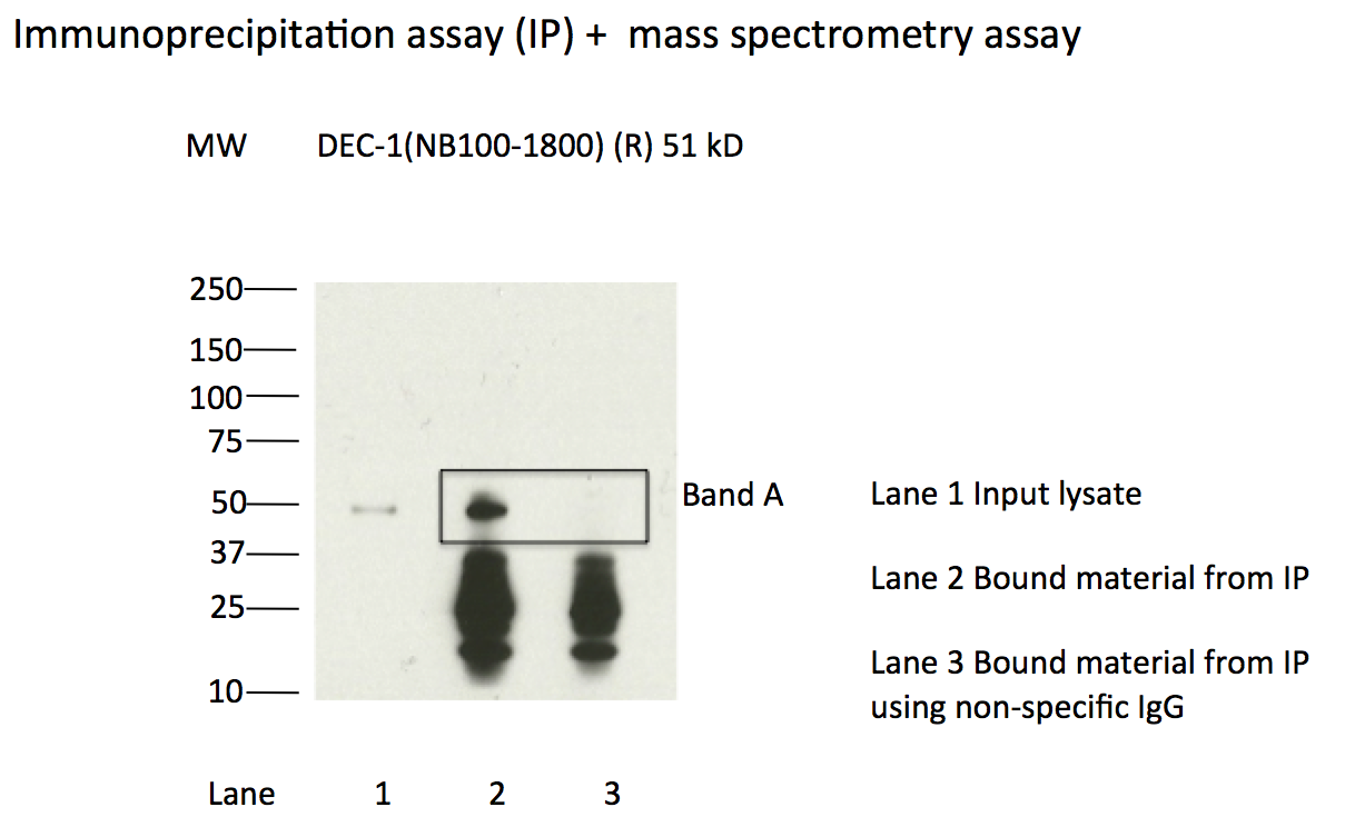
- Caption
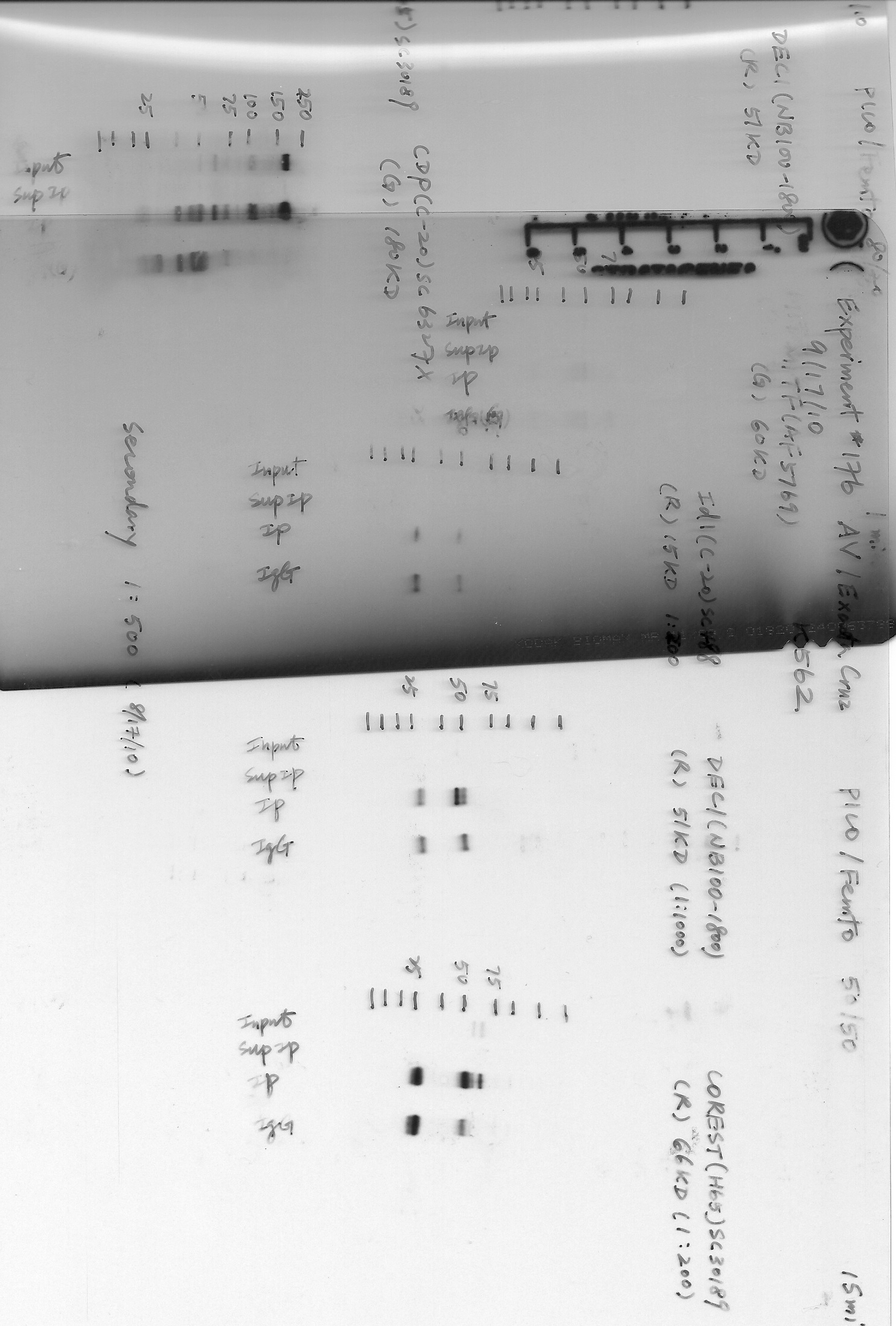
- Immunoprecipitation of DEC-1 from K562 cells using NB100-1800. Lane 1: input nuclear lysate. Lane 2: material immunoprecipitated with NB100-1800. Lane 3: material immunoprecipitated using control IgG. Band A was excised from gel and subject to analysis by mass spectrometry. Expected band size ~51kD.
- Submitted by
- Kathrina Onate
- Lab
- Michael Snyder, Stanford
- Grant
- U54HG004558
- Download
- DEC-1_IP.png
BHLHE40 (Homo sapiens)
Method: immunoprecipitation followed by mass spectrometry
compliant
- Caption
- IP followed by mass spectrometry: Briefly, protein was immunoprecipitated from K562 whole cell lysates using NB100-1800, and the IP fraction was loaded on a 10% polyacrylamide gel (NuPAGE Bis-Tris Gel) and separated with an Invitrogen NuPAGE electrophoresis system. The gel was silver-stained, gel fragments corresponding to the bands indicated were excised and destained using the SilverSNAP Stain for Mass Spectrometry (Pierce). Then proteins were treated with trypsin using the in-gel digestion method. Digested proteins were analyzed on a LTQ-Orbitrap (Thermo Scientific) b the nanoLC-ESI-MS/MS technique. Peptides were identified by the SEQUEST algorithm and filtered with a high confidence threshold (protein false discovery rate <1%, 2 peptides per protein minimum).
- Submitted by
- Kathrina Onate
- Lab
- Michael Snyder, Stanford
- Grant
- U54HG004558
- Download
- DEC1_AEK.pdf
BHLHE40 (Homo sapiens)
HEK293T
Method: immunoprecipitation
compliant
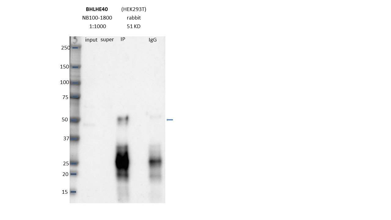
- Caption
- Immunoprecipitation was performed on nuclear extracts from the cell line: HEK293T using the antibody NB100-1800. The image shows western blot analysis of input, flowthrough, immunoprecipitate, and mock immunoprecipitate using IgG. Target molecular weight: 51.0.
- Submitted by
- Nathaniel Watson
- Lab
- Michael Snyder, Stanford
- Grant
- U54HG006996
- Download
- Expt1038_43-BHLHE40-NB100-1800.JPG
BHLHE40 (Homo sapiens)
Method: immunoprecipitation
not reviewed
- Caption
- A. Western blots on nuclear lysates from cell lines GM12878 (Lane1), K562 (Lane2), HeLaS3 (Lane3), and HepG2 (Lane4). B. Immunoprecipitation was performed on nuclear lysates from K562 cells using antibody NB100-1800 against Dec1. Lane1: Nuclear lysate. Lane 2: Unbound material from immunoprecipitation with NB100-1800. Lane 3: Bound material from immunoprecipitation with NB100-1800. Lane 4: Bound material from control immunoprecipitation with rabbit IgG. Arrow indicates band of expected size (18kD) that is highly enriched in the specifically immunoprecipitated fraction. Bands indicated by * in K562 immunoprecipitate are IgG heavy and light chains. A unique band of ~49kD is detected by Western blotting with NB100-1800 in multiple human cell lines and immunoprecipitation from K562 nuclear lysate efficiently enriches a single protein of ~49KD. Based on these observations, this antibody meets this ENCODE criterion.
- Submitted by
- Michael Snyder
- Lab
- Michael Snyder, Stanford
- Grant
- U54HG004558
BHLHE40 (Homo sapiens)
Method: immunoprecipitation followed by mass spectrometry
not reviewed
- Caption
- Immunoprecipitation of DEC1 from K562 cells using NB100-1800. Lane 1: input nuclear lysate, Lane 2: material immunoprecipitated with NB100-1800, Lane 3: material immunoprecipitated using control IgG. Bands A was excised from the gel and subject to analysis by mass spectrometry. IP followed by mass spectrometry: Briefly, protein was immunoprecipitated from K562 whole cell lysates using NB100-1800, and the IP fraction was loaded on a 10% polyacrylamide gel (NuPAGE Bis-Tris Gel) and separated with an Invitrogen NuPAGE electrophoresis system. The gel was silver-stained, gel fragments corresponding to the bands indicated were excised and destained using the SilverSNAP Stain for Mass Spectrometry (Pierce). Then proteins were trypsinized using the in-gel digestion method. Digested proteins were analyzed on an LTQ-Orbitrap (Thermo Scientific) by the nanoLC-ESI-MS/MS technique. Peptides were identified by the SEQUEST algorithm and filtered with a high confidence threshold (Protein false discovery rate < 1%, 2 peptides per protein minimum). We report 12 proteins identified in band A, although 3 of these are also present in a control immunoprecipitation and are thus likely to present due to non-specific association with the IP matrix. Of the specifically immunoprecipitated proteins, DEC1 is the most abundant (42 peptides). Based on these observations, this band is likely due to the presence of immunoprecipitated DEC1 and NB100-1800 meets the ENCODE standard for validation by this criterion.
- Submitted by
- Michael Snyder
- Lab
- Michael Snyder, Stanford
- Grant
- U54HG004558
BHLHE40 (Mus musculus)
Method: immunoprecipitation
not reviewed
- Caption
- Immunoprecipitation of CH12 and MEL nuclear extracts using anti-Dec1 antibody (NB100-1800) enriched a band of the expected molecular weight of BHLEH40 (Dec1) (~50 kD).
- Submitted by
- Michael Snyder
- Lab
- Michael Snyder, Stanford
- Grant
- RC2HG005602
BHLHE40 (Mus musculus)
Method: immunoprecipitation followed by mass spectrometry
not reviewed
- Caption
- This antibody has been validated by IP-Mass- Spec for human cell lines. Please see the validation document submitted for human cell lines for details.
- Submitted by
- Michael Snyder
- Lab
- Michael Snyder, Stanford
- Grant
- RC2HG005602