ENCAB910DVS
Antibody against Homo sapiens SRSF7
Homo sapiens
K562, HepG2
characterized to standards
Homo sapiens
any cell type or tissue
partially characterized
- Status
- released
- Source (vendor)
- MBLI
- Product ID
- RN079PW
- Lot ID
- 001
- Characterized targets
- SRSF7 (Homo sapiens)
- Host
- rabbit
- Clonality
- polyclonal
- Purification
- affinity
- Antigen description
- Peptide, N-terminus of human SRSF7
- Aliases
- xiang-dong-fu:SRSF7
- External resources
Characterizations
SRSF7 (Homo sapiens)
HepG2
Method: immunoprecipitation
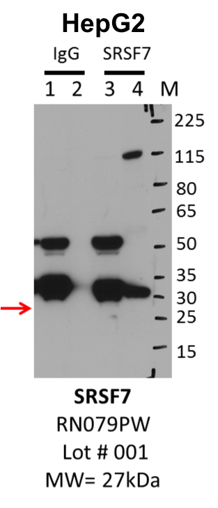
compliant
- Caption
- IP-Western Blot analysis of HepG2 whole cell lysate using SRSF7 specific antibody. Lane 1 is 1% of twenty million whole cell lysate input and lane 2 is 25% of IP enrichment using rabbit normal IgG (lanes under 'IgG'). Lane 3 is 1% of twenty million whole cell lysate input and lane 4 is 10% IP enrichment using rabbit polyclonal anti-SRSF7 antibody (lanes under 'SRSF7').
- Submitted by
- Steven Blue
- Lab
- Gene Yeo, UCSD
- Grant
- U54HG007005
- Download
- HepG2_MBLI_RN079PW_001_SRSF7.png
SRSF7 (Homo sapiens)
K562
Method: immunoprecipitation
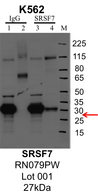
compliant
- Caption
- IP-Western Blot analysis of K562 whole cell lysate using SRSF7 specific antibody. Lane 1 is 1% of twenty million whole cell lysate input and lane 2 is 25% of IP enrichment using rabbit normal IgG (lanes under 'IgG'). Lane 3 is 1% of twenty million whole cell lysate input and lane 4 is 10% IP enrichment using rabbit polyclonal anti-SRSF7 antibody (lanes under 'SRSF7').
- Submitted by
- Steven Blue
- Lab
- Gene Yeo, UCSD
- Grant
- U54HG007005
- Download
- K562_MBLI_RN079PW_001_SRSF7.png
SRSF7 (Homo sapiens)
Method: immunoprecipitation
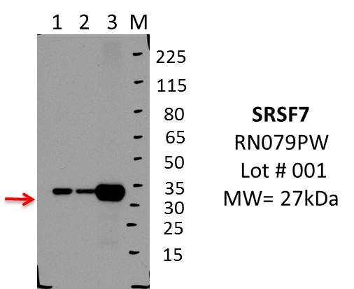
not submitted for review by lab
- Caption
- IP-WB analysis of MCF7 whole cell lysate using SRSF7 specific antibody. Lane 1 is 2.5% of 0.5mg input lysate, lane 2 is 2.5% of supernatant after immunoprecipitation and Lane 3 is 50% of IP enrichment using rabbit polyclonal Anti-SRSF7(9G8)pAb. This antibody passes preliminary validation and will be further pursued for primary and secondary validation.
- Submitted by
- Balaji Sundararaman
- Lab
- Gene Yeo, UCSD
- Grant
- U54HG007005
- Download
- MBLI_RN079PW_001_SRSF7.jpg
SRSF7 (Homo sapiens)
Method: immunoprecipitation
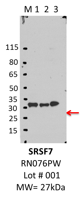
not submitted for review by lab
- Caption
- IP-WB characterization of SRSF7 specific antibody in K562 cell line . Lane 1 is 2.5% of five million K562 whole cell lysate Input, lane 2 is 2.5% of supernatant after immunoprecipitation and Lane 3 is 50% of IP enrichment using rabbit polyclonal Anti-SRSF7(9G8)pAb. This antibody passes preliminary validation and will be further pursued for primary and secondary validation.
- Submitted by
- Balaji Sundararaman
- Lab
- Gene Yeo, UCSD
- Grant
- U54HG007005
- Download
- MBLI_RN079PW_001_SRSF7_K562.png
SRSF7 (Homo sapiens)
Method: knockdown or knockout
compliant
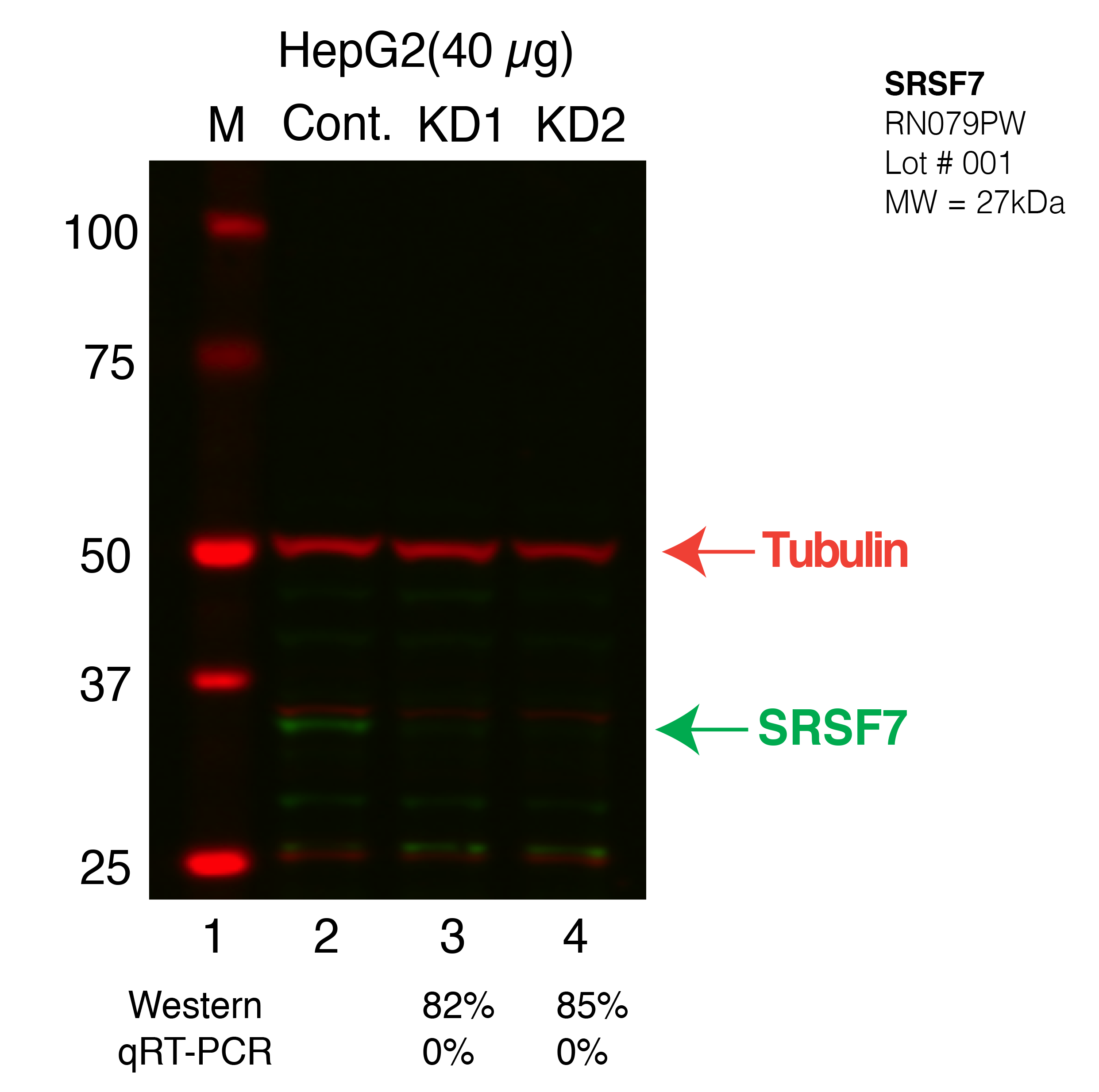
- Caption
- Western blot following CRISPR against SRSF7 in HepG2 whole cell lysate using SRSF7 specific antibody. Lane 1 is a ladder, lane 2 is HepG2 non-targeting control knockdown, lane 3 and 4 are two different CRISPR against SRSF7. SRSF7 protein appears as the green band, Tubulin serves as a control and appears in red.
- Submitted by
- Xintao Wei
- Lab
- Brenton Graveley, UConn
- Grant
- U54HG007005
- Download
- SRSF7-HEPG2-CRISPR.png
SRSF7 (Homo sapiens)
Method: knockdown or knockout
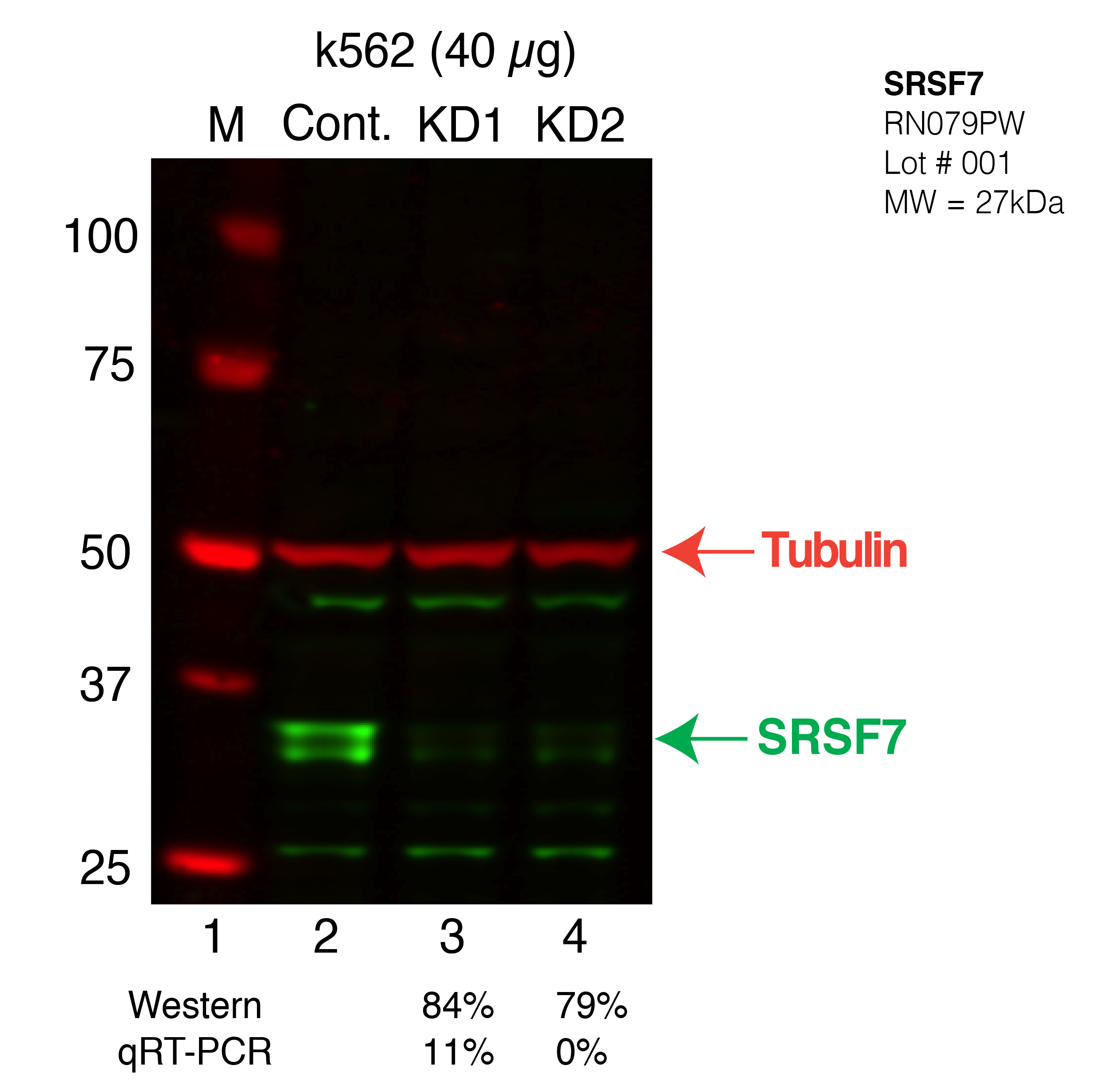
compliant
- Caption
- Western blot following CRISPR against SRSF7 in k562 whole cell lysate using SRSF7 specific antibody. Lane 1 is a ladder, lane 2 is k562 non-targeting control knockdown, lane 3 and 4 are two different CRISPR against SRSF7. SRSF7 protein appears as the green band, Tubulin serves as a control and appears in red.
- Submitted by
- Xintao Wei
- Lab
- Brenton Graveley, UConn
- Grant
- U54HG007005
- Download
- SRSF7-K562-CRISPR.png
SRSF7 (Homo sapiens)
Method: knockdown or knockout
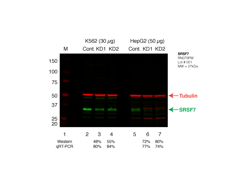
compliant
- Caption
- Western blot following shRNA against SRSF7 in K562 and HepG2 whole cell lysate using SRSF7 specific antibody. Lane 1 is a ladder, lane 2 is K562 non-targeting control knockdown, lane 2 and 3 are two different shRNAs against SRSF7. Lanes 5-7 follow the same pattern, but in HepG2. SRSF7 protein appears as the green band, GAPDH serves as a control and appears in red.
- Submitted by
- Brenton Graveley
- Lab
- Brenton Graveley, UConn
- Grant
- U54HG007005
- Download
- SRSF7_Secondary_Western.png