ENCAB027LVC
Antibody against Homo sapiens SNIP1
Homo sapiens
K562, MCF-7, HepG2, HEK293T
characterized to standards
Homo sapiens
any cell type or tissue
partially characterized
- Status
- released
- Source (vendor)
- Bethyl Labs
- Product ID
- A300-371A
- Lot ID
- 1
- Characterized targets
- SNIP1 (Homo sapiens)
- Host
- rabbit
- Clonality
- polyclonal
- Purification
- affinity
- Isotype
- IgG
- External resources
Characterizations
SNIP1 (Homo sapiens)
MCF-7
Method: immunoprecipitation
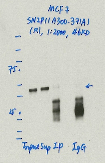
compliant
- Caption
- Immunoprecipitation was performed on nuclear extracts from the cell line: MCF-7, using the antibody A300-371A. The blot shows western blot analysis of input, flowthrough, immunoprecipitate and mock immunoprecipitate using IgG.Molecular Weight: 45.778
- Reviewer comment
- Though there is nothing explicit in the ENCODE standards about the strength of immunoreactive signal in the IP relative to that of the input and supernatant, it is a bit concerning that it is far stronger in both those lanes relative to the IP.
- Submitted by
- Denis Salins
- Lab
- Michael Snyder, Stanford
- Grant
- U54HG006996
- Download
- 1062_3_SNIP1_A300-371A_MCF7.jpg
SNIP1 (Homo sapiens)
HepG2
Method: immunoprecipitation
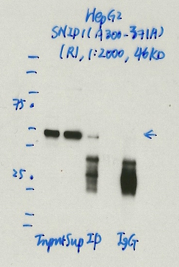
compliant
- Caption
- Immunoprecipitation was performed on nuclear extracts from the cell line: HepG2, using the antibody A300-371A. The blot shows western blot analysis of input, flowthrough, immunoprecipitate and mock immunoprecipitate using IgG.Molecular Weight: 45.778
- Reviewer comment
- Though there is nothing explicit in the ENCODE standards about the strength of immunoreactive signal in the IP relative to that of the input and supernatant, it is a bit concerning that it is far stronger in both those lanes relative to the IP.
- Submitted by
- Denis Salins
- Lab
- Michael Snyder, Stanford
- Grant
- U54HG006996
- Download
- 1062_4_SNIP1_A300-371A_HepG2.jpg
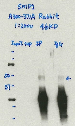
SNIP1 (Homo sapiens)
K562
Method: immunoprecipitation
compliant
- Caption
- Immunoprecipitation was performed on nuclear extracts from the cell line: K562, using the antibody A300-371A. The blot shows western blot analysis of input, flowthrough, immunoprecipitate and mock immunoprecipitate using IgG.
- Submitted by
- Denis Salins
- Lab
- Michael Snyder, Stanford
- Grant
- U54HG006996
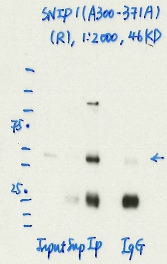
SNIP1 (Homo sapiens)
Method: immunoprecipitation
not submitted for review by lab
- Caption
- IP-WB analysis of K562 whole cell lysate using SNIP1 specific antibody. Lane 1 is 2.5% of 0.5mg input lysate, lane 2 is 2.5% of supernatant after immunoprecipitation and Lane 3 is 50% of IP enrichment using rabbit polyclonal SNIP1 antibody. This antibody did not meet our primary validation criteria using our standard IP protocol in the indicated cell type.
- Submitted by
- Balaji Sundararaman
- Lab
- Gene Yeo, UCSD
- Grant
- U54HG007005
- Download
- Bethyl_A300-371A_1_SNIP1.png
SNIP1 (Homo sapiens)
Method: immunoprecipitation
not submitted for review by lab
- Caption
- Immunoprecipitation was performed on nuclear extracts from the cell line: GM12878, using the antibody A300-371A. The blot shows western blot analysis of input, flowthrough, immunoprecipitate and mock immunoprecipitate using IgG.Molecular Weight: 45.778
- Submitted by
- Denis Salins
- Lab
- Michael Snyder, Stanford
- Grant
- U54HG006996
- Download
- expt1063_4-SNIP1-A300-371A.jpg
SNIP1 (Homo sapiens)
HEK293T
Method: immunoprecipitation
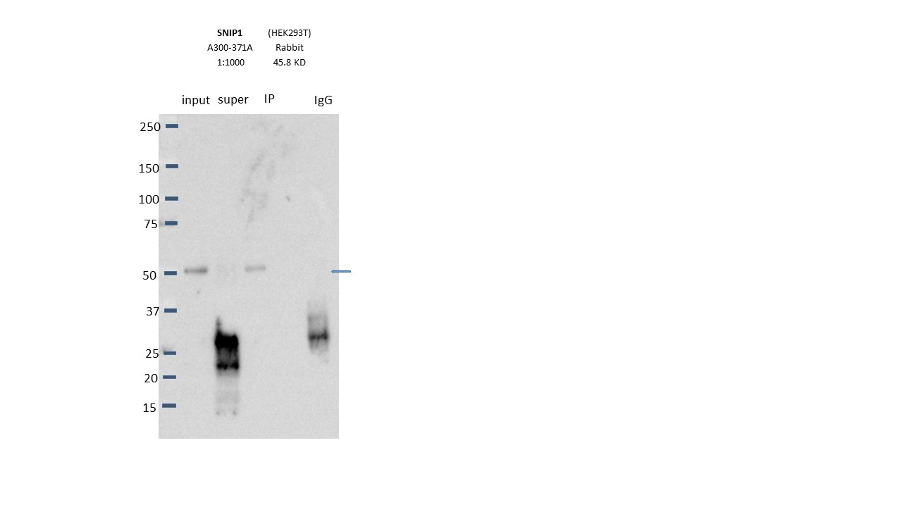
compliant
- Caption
- Immunoprecipitation was performed on nuclear extracts from the cell line: HEK293T, using the antibody A300-371A. The blot shows western blot analysis of input, flowthrough, immunoprecipitate and mock immunoprecipitate using IgG.Molecular Weight: 45.778
- Submitted by
- Nathaniel Watson
- Lab
- Michael Snyder, Stanford
- Grant
- U54HG006996
- Download
- Expt1113_3-SNIP1-A300-371A.jpg
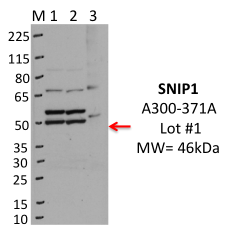
SNIP1 (Homo sapiens)
K562
Method: immunoprecipitation
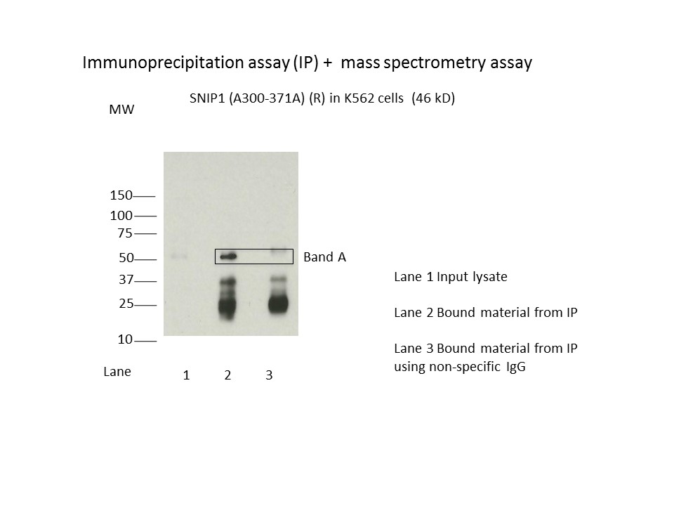
compliant
- Caption
- Immunoprecipitation was performed on nuclear extracts from the cell line K562 using the antibody A300-371A. Lane 1: input nuclear lysate. Lane 2: material immunoprecipitated with antibody. Lane 3: material immunoprecipitated using control IgG. Marked bands were excised from gel and subjected to analysis by mass spectrometry. Target molecular weight: 45.778.
- Submitted by
- Nathaniel Watson
- Lab
- Michael Snyder, Stanford
- Grant
- U54HG006996
- Download
- SNIP1.jpeg
SNIP1 (Homo sapiens)
Method: immunoprecipitation followed by mass spectrometry
compliant
- Caption
- IP followed by mass spectrometry. Briefly, protein was immunoprecipitated from K562 nuclear cell lysates using the antibody A300-371A, and the IP fraction was loaded on a 10% polyacrylamide gel (NuPAGEBis-Tris Gel) and separated with an Invitrogen NuPAGE electrophoresis system. The gel was stained by ColloidialCoomassie G-250 stain, gel fragments corresponding to the bands indicated were excised. Then proteins were trypsinized using the in-gel digestion method. Digested proteins were analyzed on an Orbitrap Elite mass spectrometer (Thermo Scientific) by the nanoLC-ESI-MS/MS technique. Peptides were identified by the SEQUEST algorithm and filtered with a high confidence threshold (Peptide false discovery rate < 1%, 2 unique peptides per protein minimum, mass error < 10 ppm).
- Submitter comment
- None of the proteins ranked above or equal to SNIP1 are sequence-specific TFs.
- Submitted by
- Nathaniel Watson
- Lab
- Michael Snyder, Stanford
- Grant
- U54HG006996
- Download
- SNIP1_A300-371A final.pdf